Avian Intestines - Anatomy & Physiology
Structure
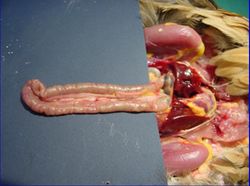
The intestines occupy the caudal part of the body. They contact the reproductive organs and the gizzard. The small intestine is long and relatively uniform in shape and size. There is no demarcation between the jejunum and the ileum.
The duodenum passes caudally over the gizzard then loops back towards the stomach where it joins the jejunum. It arises from the right dorsal aspect of the gizzard. The loop lies ventral on the abdominal floor and the pancreas lies within the loops. Three pancreatic ducts and one bile duct enter the caudal duodenum at a common papilla.
Jejunum
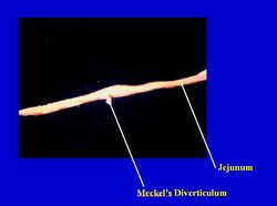
The jejunum has loose coils around the mesentery. It has thin walls so its content appears green. It is suspended from the dorsal wall of the abdomen by the mesentery.
Ileum
The ileum begins opposite the apices of the caeca or at the vitelline diverticula. It is suspended from the dorsal wall of the abdomen by the mesentery .
colon
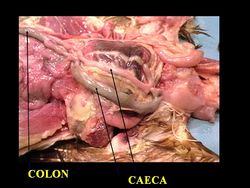
The short colon lies ventral to the synsacrum and opens into the cloaca. It runs ventral to the vertebrae and terminates in the coprodeum. Amino acids and glucose can be absorbed here.
Two caeca from the ileocaecal junction run with the ileum caudally. They are blind sacs. They extend towards the liver then fold back on themselves. The mesentery runs between the caeca then on towards the ileum. It often contains dark coloured material. There are three parts of each caecum. It is where the bacterial breakdown of cellulose occurs. Antiperistaltic movements transport chyme and the caeca are emptied a few times per day. Unlike mammals, there are no lacteals in the epithelium.
Vitelline Diverticula
The vitelline diverticulum is a small outgrowth on the jejunum. It is the former connection with the yolk sac. It is also called Meckel's diverticulum.
Function
Vasculature
Innervation
Lymphatics
Patches of lymphoid nodules are present in Peyer's Patches. They are most abundant in the duodenum. There are no mesenteric lymph nodes.
Histology
The serous coat has nerve plexuses. There are columnar epithelium and goblet cells, with smooth muscle in folds at the base. The caecal sphincter at the proximal part contains a lot of lymphoid tissue (caecal tonsil). The middle section has thin walls and appears green and the bulbous blind ends have thicker walls.
For more information, see: small intestine histology.
Species Differences
The duck and goose have several loops of 'U' shaped jejunum. Pigeons have a circular mass of jejunum with inner and outer turns. Long caeca are present in the turkey and chicken. Pigeons and song birds have short caeca. Parrots do not have caeca. The dorsal and ventral lobes of the pancreas are connected dorsally in poultry.
Links
Click here for information on the Small Intestine - Anatomy & Physiology
Click here for more information on the Large Intestine - Anatomy & Physiology
| Avian Intestines - Anatomy & Physiology Learning Resources | |
|---|---|
 Test your knowledge using flashcard type questions |
Avian Alimentary Tract |
Error in widget FBRecommend: unable to write file /var/www/wikivet.net/extensions/Widgets/compiled_templates/wrt699f9a4f50bd81_44676286 Error in widget google+: unable to write file /var/www/wikivet.net/extensions/Widgets/compiled_templates/wrt699f9a4f605aa4_09193051 Error in widget TwitterTweet: unable to write file /var/www/wikivet.net/extensions/Widgets/compiled_templates/wrt699f9a4f70fb05_20896481
|
| WikiVet® Introduction - Help WikiVet - Report a Problem |


