Difference between revisions of "B cell differentiation"
| (57 intermediate revisions by 6 users not shown) | |||
| Line 1: | Line 1: | ||
| − | < | + | {{OpenPagesTop}} |
| − | < | + | [[Image:B-cell immunity.jpg|thumb|right|250pxl|B-cell differentiation - B. Catchpole, RVC 2008]] |
| − | + | ==Introduction== | |
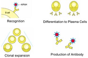
| + | <p>Mature [[B cells]] that undergo stimulation by an antigen undergo class switching, and differentiate into either '''plasma''' or '''memory''' cells. </p> | ||
| + | <p>In the paracortex region of the [[Lymph Nodes - Anatomy & Physiology|lymph node]] binding to [[Major_Histocompatability_Complexes#MHC_II|MHC II]] in the presence of IL-4 produced by the [[Lymphocytes#Helper CD4+|CD4<sup>+</sup> T cells]] (T<sub>H</sub>2 type) causes the B cells to differentiate; most will become plasma cells, however a small number will become memory cells.</p> | ||
| − | + | Follicular dendritic cells present in the germinal centers of peripheral lymphoid organs can absorb intact antigen onto their surface to present to B cells to stimulate differentiation. | |
| − | |||
| − | |||
| − | |||
| − | |||
| − | Follicular dendritic | ||
==Plasma cells== | ==Plasma cells== | ||
===Appearance=== | ===Appearance=== | ||
| − | <p>Plasma cells are oval, around 9µm and have a round prominent nucleus. The cytoplasm is extensive and | + | <p>Plasma cells are oval, around 9µm and have a round prominent nucleus. The cytoplasm is extensive and strongly basophilic when stained. It contains large amounts of rough endoplasmic reticulum and the Golgi apparatus is large and appears as a clear crescent-shaped structure near the nucleus. Some plasma cells, known as '''"Mott cells"''', accumulate considerable quantities of, perhaps abnormal, antibody and this appears as a large eosinophilic blob filling the cytoplasm and displacing the nucleus to one side. These blobs are called '''"Russell Bodies"'''.</p> |
| + | |||
===Function=== | ===Function=== | ||
| − | <p>Plasma cells produce immunoglobulins/antibodies (thousands a second). The immunoglobulin binding specificity is identical to the binding specificity of the BCR on the [[ | + | [[Image:B-cell-Ab.jpg|thumb|right|150px|Plasma cell antibody production - B. Catchpole, RVC 2008]] |
| + | <p>Plasma cells produce immunoglobulins/antibodies (thousands a second). The immunoglobulin binding specificity is identical to the binding specificity of the B Cell receptor (BCR) on the [[B cells|B cell]] that the cell has differentiated from. This means that when a B cell has a BCR that can effectively bind to an antigen the immunoglobulins produced by the plasma cell can bind to that antigen.</p> | ||
<p>Although they can live for months most plasma cells only live for a few days and do not replicate in this time.</p> | <p>Although they can live for months most plasma cells only live for a few days and do not replicate in this time.</p> | ||
| − | Interaction of a B-cell with antigen results in clonal expansion, as does activation by T-cells. The majority of B-cell clones mature into plasma cells. Plasma cells are found in the splenic [[Spleen - Anatomy & Physiology#Red Pulp|red pulp]], [[Lymph Nodes - Anatomy & Physiology#Lymph Nodes|lymph node medulla]] and [[Bone Marrow - Anatomy & Physiology|bone marrow]]. Plasma cells are the terminal differentiation state of B-cells. They migrate to the medullary cords where their | + | Interaction of a B-cell with antigen results in clonal expansion, as does activation by T-cells. The majority of B-cell clones mature into plasma cells. Plasma cells are found in the splenic [[Spleen - Anatomy & Physiology#Red Pulp|red pulp]], [[Lymph Nodes - Anatomy & Physiology#Lymph Nodes|lymph node medulla]] and [[Bone Marrow - Anatomy & Physiology|bone marrow]]. Plasma cells are the terminal differentiation state of B-cells. They migrate to the medullary cords where their sole function is to secrete antibody. |
| + | |||
===Class switching=== | ===Class switching=== | ||
| − | <p>Initially plasma cell produce [[Immunoglobulin M | + | <p>Initially plasma cell produce [[Immunoglobulin M|Immunoglobulin M (IgM)]] however this is not always the most appropriate Ig to be produced and therefore stimulation by [[Lymphocytes#Helper CD4+|T cells]] and interleukins causes the plasma cells to undergo class switching to produce different classes of Immunoglobulins. |
| − | *In mucosal B cells plasma cells CD40 interaction (with | + | *In mucosal B cells plasma cells CD40 interaction (with T<sub>H</sub>2 CD40L) and Il-10 stimulates class switching to [[Immunoglobulin A|IgA]] |
| − | *[[Eosinophils | + | *[[Eosinophils|Eosinophils]] produce Il-13 which promotes class switching to [[Immunoglobulin E|IgE]]</p> |
| − | <p>Plasma cells produced in the first immune response to an antigen are mainly of the class IgM whereas those produced from memory cells in the second immune response are mainly of the [[Immunoglobulin G | + | <p>Plasma cells produced in the first immune response to an antigen are mainly of the class IgM whereas those produced from memory cells in the second immune response are mainly of the [[Immunoglobulin G|IgG]] class.</p> |
| − | |||
==T-Cell Dependent and Independent Responses== | ==T-Cell Dependent and Independent Responses== | ||
| − | There are two types of B cell response to antigen | + | There are two types of B cell response to antigen; one is dependant and the other independant of T cell interaction. |
===T-Cell Independent Response=== | ===T-Cell Independent Response=== | ||
| − | [[Image:T Cell Independent Diagram.jpg|right|thumb| | + | [[Image:T Cell Independent Diagram.jpg|right|thumb|200px|Diagram of the T Cell Independent Repsonse - Copyright nabrown RVC]] |
| − | + | B cells are activated in this instance by '''non-protinaceous antigens''' such as lipopolysaccharides which act as mitogens for the B cells by stimulating cell division. | |
| − | + | These antigens only stimulate '''primary''' immune responses (production of [[Immunoglobulin M|IgM]]) and do not produce memory cells. Initial and subsequent exposure to an antigen stimulates an identical response from the B cell. | |
| − | |||
| − | |||
| − | |||
| − | |||
| − | |||
===T-Cell Dependent Response=== | ===T-Cell Dependent Response=== | ||
| − | [[Image:T cell Dependent Response Diagram.jpg|thumb|right| | + | [[Image:T cell Dependent Response Diagram.jpg|thumb|right|200px|Diagram showing the T cell dependent response (primary and secondary phases) - Copyright nabrown RVC]] |
| − | + | The B cell response is to '''protinaceous antigens''' and T cells moderate the response, which occurs in the germinal centres of follicles in secondary lymphoid tissues. | |
| − | + | ||
| − | + | B cells then act as antigen presenting cells on [[Major Histocompatability Complexes#Structure and Function Of MHC Class II|MHC II]], with T cells interacting via cell surface receptors: | |
| − | + | *CD40L on the T cell | |
| − | + | *CD40 on the B cell | |
| − | + | T cells produce [[Cytokines|'''cytokines''']] such as IL-4 which induces [[Immunoglobulins_-_Overview#Immunoglobulin_Class_Switching|class switching]] from IgM and the formation of memory cells. | |
| − | |||
| − | |||
| − | |||
| − | |||
| − | |||
====Primary T Cell Dependent Response==== | ====Primary T Cell Dependent Response==== | ||
| − | + | The first exposure of an individual to a particular antigen is referred to as '''priming'''. The measurable antibody response is called the '''primary immune response'''. The delay of 5-7 days before antibody is produced is called the '''Lag Phase''' during which time the B cells undergo clonal expansion and form plasma cells. IgM antibody is produced first and will begin to appear in the blood; this stage is called the '''Log Phase'''. | |
| − | + | ||
| − | + | The log phase will peak after about 10-14 days and the '''Plateau Phase''' will then occur. | |
| − | + | Class switching occurs replacing decreasing levels of [[Immunoglobulin M|IgM]] with [[Immunoglobulin G|IgG]]. Antibody levels then begin to decline as plasma cells undergo apoptosis. | |
| − | + | ||
| − | + | *After primary immunisation, it usually takes around 7-10 days for a measurable antibody response to become detectable. This latent period is the time necessary for the antigen to contact specific cells, for the cells involved to interact and expand, and for plasma cells to secrete antibody. | |
| − | + | During the primary response, [[Immunoglobulin G|IgG]] production lags about a week behind that of [[Immunoglobulin M|IgM]]. | |
| − | |||
| − | |||
| − | *After primary immunisation, it usually takes around 7-10 days for a measurable antibody response to become detectable | ||
| − | |||
| − | |||
| − | |||
| − | |||
====Secondary T Cell Dependent Response==== | ====Secondary T Cell Dependent Response==== | ||
| − | + | The production of antibody to any antigen ceases within a few weeks of immunisation as the antigen disappears from the body; However, the animal retains '''immunological memory''' of the antigen which occurs after the primary response - it is seen on the second and subsequent exposures to the same antigen. In effect, there is an expanded pool of memory B and T cells from the first exposure to the particular antigen which permits a shorter lag phase and a longer plateau phase as antibody persists for an extended time. There is a higher antibody titre overall due to cloanal expansion and '''Long-lived memory cells''' which are qualitatively distinct are produced. Antigen is retained within the immune system on the surface of follicular dendritic cells and this can stimulate the immune system for years. | |
| − | |||
| − | |||
| − | |||
| − | |||
| − | |||
| − | |||
| − | |||
| − | |||
| − | |||
| − | |||
| − | |||
| − | |||
| + | The level of [[Immunoglobulin M|IgM]] antibody production in the secondary response is similar to the primary response; the increase in antibody production occurs with [[Immunoglobulin G|IgG]]. | ||
| − | === | + | ===Plasma Cell Pathology=== |
| − | + | If many plasma cells are present in blood, it is due to a plasma cell tumour that has begun to metastasise. | |
==Memory cells== | ==Memory cells== | ||
| − | <p>The differentiated cells that remain in the cortex become memory cells and these proliferate and form germinal centres in the [[Lymph Nodes - Anatomy & Physiology|lymph node]].</p> | + | <p>The differentiated B cells that remain in the cortex become memory cells and these proliferate and form germinal centres in the [[Lymph Nodes - Anatomy & Physiology|lymph node]].</p> |
<p>Memory cells are long lived and responsible for long term immunity providing the immune system with a memory of previously encountered antigens. When they experience an antigen again they proliferate and differentiate into plasma cells. This response produces up to ten times more plasma cells than the original exposure to the antigen and is why the second immune response to an antigen is both more rapid and much stronger than the first response.</p> | <p>Memory cells are long lived and responsible for long term immunity providing the immune system with a memory of previously encountered antigens. When they experience an antigen again they proliferate and differentiate into plasma cells. This response produces up to ten times more plasma cells than the original exposure to the antigen and is why the second immune response to an antigen is both more rapid and much stronger than the first response.</p> | ||
| − | + | <br><br> | |
| − | + | {{Jim Bee 2007}} | |
| − | |||
| − | |||
| − | |||
| − | |||
| − | + | {{OpenPages}} | |
| − | + | [[Category:Lymphocytes|C]] | |
| − | + | [[Category:Adaptive Immune System]] | |
Latest revision as of 16:17, 2 July 2012
Introduction
Mature B cells that undergo stimulation by an antigen undergo class switching, and differentiate into either plasma or memory cells.
In the paracortex region of the lymph node binding to MHC II in the presence of IL-4 produced by the CD4+ T cells (TH2 type) causes the B cells to differentiate; most will become plasma cells, however a small number will become memory cells.
Follicular dendritic cells present in the germinal centers of peripheral lymphoid organs can absorb intact antigen onto their surface to present to B cells to stimulate differentiation.
Plasma cells
Appearance
Plasma cells are oval, around 9µm and have a round prominent nucleus. The cytoplasm is extensive and strongly basophilic when stained. It contains large amounts of rough endoplasmic reticulum and the Golgi apparatus is large and appears as a clear crescent-shaped structure near the nucleus. Some plasma cells, known as "Mott cells", accumulate considerable quantities of, perhaps abnormal, antibody and this appears as a large eosinophilic blob filling the cytoplasm and displacing the nucleus to one side. These blobs are called "Russell Bodies".
Function
Plasma cells produce immunoglobulins/antibodies (thousands a second). The immunoglobulin binding specificity is identical to the binding specificity of the B Cell receptor (BCR) on the B cell that the cell has differentiated from. This means that when a B cell has a BCR that can effectively bind to an antigen the immunoglobulins produced by the plasma cell can bind to that antigen.
Although they can live for months most plasma cells only live for a few days and do not replicate in this time.
Interaction of a B-cell with antigen results in clonal expansion, as does activation by T-cells. The majority of B-cell clones mature into plasma cells. Plasma cells are found in the splenic red pulp, lymph node medulla and bone marrow. Plasma cells are the terminal differentiation state of B-cells. They migrate to the medullary cords where their sole function is to secrete antibody.
Class switching
Initially plasma cell produce Immunoglobulin M (IgM) however this is not always the most appropriate Ig to be produced and therefore stimulation by T cells and interleukins causes the plasma cells to undergo class switching to produce different classes of Immunoglobulins.
- In mucosal B cells plasma cells CD40 interaction (with TH2 CD40L) and Il-10 stimulates class switching to IgA
- Eosinophils produce Il-13 which promotes class switching to IgE
Plasma cells produced in the first immune response to an antigen are mainly of the class IgM whereas those produced from memory cells in the second immune response are mainly of the IgG class.
T-Cell Dependent and Independent Responses
There are two types of B cell response to antigen; one is dependant and the other independant of T cell interaction.
T-Cell Independent Response
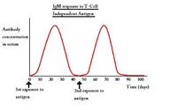
B cells are activated in this instance by non-protinaceous antigens such as lipopolysaccharides which act as mitogens for the B cells by stimulating cell division. These antigens only stimulate primary immune responses (production of IgM) and do not produce memory cells. Initial and subsequent exposure to an antigen stimulates an identical response from the B cell.
T-Cell Dependent Response
The B cell response is to protinaceous antigens and T cells moderate the response, which occurs in the germinal centres of follicles in secondary lymphoid tissues.
B cells then act as antigen presenting cells on MHC II, with T cells interacting via cell surface receptors:
- CD40L on the T cell
- CD40 on the B cell
T cells produce cytokines such as IL-4 which induces class switching from IgM and the formation of memory cells.
Primary T Cell Dependent Response
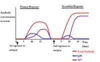
The first exposure of an individual to a particular antigen is referred to as priming. The measurable antibody response is called the primary immune response. The delay of 5-7 days before antibody is produced is called the Lag Phase during which time the B cells undergo clonal expansion and form plasma cells. IgM antibody is produced first and will begin to appear in the blood; this stage is called the Log Phase.
The log phase will peak after about 10-14 days and the Plateau Phase will then occur. Class switching occurs replacing decreasing levels of IgM with IgG. Antibody levels then begin to decline as plasma cells undergo apoptosis.
- After primary immunisation, it usually takes around 7-10 days for a measurable antibody response to become detectable. This latent period is the time necessary for the antigen to contact specific cells, for the cells involved to interact and expand, and for plasma cells to secrete antibody.
During the primary response, IgG production lags about a week behind that of IgM.
Secondary T Cell Dependent Response
The production of antibody to any antigen ceases within a few weeks of immunisation as the antigen disappears from the body; However, the animal retains immunological memory of the antigen which occurs after the primary response - it is seen on the second and subsequent exposures to the same antigen. In effect, there is an expanded pool of memory B and T cells from the first exposure to the particular antigen which permits a shorter lag phase and a longer plateau phase as antibody persists for an extended time. There is a higher antibody titre overall due to cloanal expansion and Long-lived memory cells which are qualitatively distinct are produced. Antigen is retained within the immune system on the surface of follicular dendritic cells and this can stimulate the immune system for years.
The level of IgM antibody production in the secondary response is similar to the primary response; the increase in antibody production occurs with IgG.
Plasma Cell Pathology
If many plasma cells are present in blood, it is due to a plasma cell tumour that has begun to metastasise.
Memory cells
The differentiated B cells that remain in the cortex become memory cells and these proliferate and form germinal centres in the lymph node.
Memory cells are long lived and responsible for long term immunity providing the immune system with a memory of previously encountered antigens. When they experience an antigen again they proliferate and differentiate into plasma cells. This response produces up to ten times more plasma cells than the original exposure to the antigen and is why the second immune response to an antigen is both more rapid and much stronger than the first response.
| Originally funded by the RVC Jim Bee Award 2007 |
Error in widget FBRecommend: unable to write file /var/www/wikivet.net/extensions/Widgets/compiled_templates/wrt694a1b8bd96f92_78584871 Error in widget google+: unable to write file /var/www/wikivet.net/extensions/Widgets/compiled_templates/wrt694a1b8be39005_67373997 Error in widget TwitterTweet: unable to write file /var/www/wikivet.net/extensions/Widgets/compiled_templates/wrt694a1b8be7fa91_59745686
|
| WikiVet® Introduction - Help WikiVet - Report a Problem |


