Gastric Ulceration - Dog
| This article is still under construction. |
| Also known as: | Gastrointestinal ulceration |
| See also: | Gastric Ulceration - all species |
Description
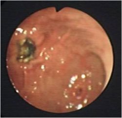
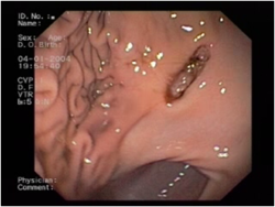
Is a round or oval punched out lesion of the gastric mucosa ranging from 1-4 cm in diameter.
There are many disease associations including:
| Disease type | E.g. |
|---|---|
| Hypotension | Shock, Sepsis |
| Drug - induced | Non-steroidal anti-inflammatory drugs (NSAIDs) |
| Idiopathic | Stress, exercise induced |
| Inflammatory | Gastritis, Pancreatitis |
| Neoplastic | Adenocarcinoma, lymphosarcoma, leiomyoma, gastrinoma (Zollinger-Ellison syndrome), |
| Metabolic/endocrine | Hypoadrenocorticism, liver disease, uraemia, Disseminated Intravascular Coagulation (DIC), mastocytosis and hypergastrinaemia |
Gastric ulceration is caused by damage to the gastric mucosa through the above mechanisms. NSAIDs directly damage the mucosa and interfere with the prostaglandin synthesis. Gastric ulceration is worsened by the use of NSAIDs in combination with corticosteroids. This risk can be minimised by using cyclooxygenase-1 (COX-1) sparing NSAIDs.
Gastric acid hypersecretion following mast cell degranulation of histamine and gastrin secretion from gastrinomas is a major cause of gastric ulceration.
Signalment
Sled dogs are prone to gastric ulceration.
Diagnosis
Clinical Signs
History may involve access to toxins and drugs such as NSAIDs. Clinical Signs can include vomiting, haematemesis, malaena, pale mucous membranes, abdominal pain, weakness, inappetance and hypersalivation which can progress to circulatory compromise.
Laboratory Tests
Haematology
Anaemia which may be regenerative initially, and can progress to microcytic, hypochromic and minutely regenerative anaemia. A thrombocytosis may also be present. If a stress leucogram (lymphopenia and neutrophilia) is not present this is supportive of hypoadrenocorticism. Examination of the buffy coat may detect mastocytosis. A Neutrophilia and a left shift are indicative of inflammation or gastric perforation. There may also be abnormalities in haemostasis.
Biochemistry
Increased liver enzymes and bilirubin, decreased urea, albumin and cholesterol will indicate hepatic disease as an underlying problem. If renal disease is present, an azotaemia will be present on biochemistry. If Hypoadrenocorticism is the cause of the ulceration, it is likely biochemistry will show a Sodium:Potassium ratio of less than 27:1. If the animal is vomiting this will lead to electrolyte and acid-base abnormalities, a metabolic alkalosis, hypokalaemia and hypochloraemia.
Urinalysis
Animals will be dehydrated resulting in Hypersthenuria. If renal disease is the underlying cause, urine may be isosthenuric.
Plain radiography
Not usually diagnostic but can rule out differentials such as foreign bodies and peritonitis.
Positive contrast radiography
May show filling defects.
Ultrasonography
Shows gastric thickening and rules out differentials.
Endoscopy and Biopsy
Diagnostic test of choice and allows biopsies to be taken. NSAID related ulcers are reguarly located in the antrum and there is limited mucosal thickening or irregularity whereas ulcerated gastric tumours will have thickened mucosa and edges. Any biopsies should be taken at the edge of normal and diseased to avoid further deepening or perforation.
Treatment
The main aim is to treat any primary underlying cause whilst giving general support. This may be hydrating, restoring electrolytes and acid-base and also helping the gastric lining to recover. Anti-ulcerative therapy should be continued for up to 6-8 weeks.
Fluid Therapy
Depends upon degree of dehydration, prescence of shock and any other diseases that are affected by volume. Prolonged vomiting or anorexia may lead to hypokalaemia so KCl may need adding to any fluids given. Normal rates for treatment of shock apply with dehydration being overcome by a fluid rate over 24 hours to replace the defecits along with a maintenance rate.
Acid-base correction
Imbalances should be corrected after taking a blood gas reading.
- If metabolic acidotic: give sodium bicarbonate but do repeated blood gas
- If metabolic alkalosis: replace volume defecit with intravenous NaCl and KCl.
- Blocking of acid secretion:
- Histamine receptor antagonists:
- cimetidine
- ranitidine
- famotidine
- Gastrin antagonists:
- proglumide
- Acetylcholine receptor antagonists:
- atropine
- pirenzepine
- Adenyl cyclase inhibitors:
- prostaglangin E2 (PGE) analogues (misoprostol)
- H+:K+ATPase inhibitors :- for use when patient is refractory to histamine antagonists
- omeprazole - good for exercise induced gastric ulceration
- Histamine receptor antagonists:
Mucosal protectants
Such as misoprostol can be given alongside NSAIDs to decrease the risk of ulceration. Sucralfate which is polyaluminium sucrose sulphate, binds to damaged mucosa and assists in the treatment of gastric ulceration. It is best given 2 hours after acid inhibitors to prevent interference.
Prophylaxis
Prophylactic treatment has been shown not to prevent gastric ulceration. Sucralfate is reported to be the best drug in patients receiving high doses of glucocorticoids.
Anti-emetics
Indicated if vomiting is severe causing fluid and electrolyte imbalances and discomfort. See Anti-emetics for drug details.
Analgesia
Is best provided by opiods such as buprenorphine, pethidine and fentanyl.
Antibiotics
Animals suffering from shock and gastric barrier dysfunction may require prophylactic antibiotic cover. First line drugs include ampicillin or a cephalosporin which are effective against Gram-positive, some Gram-negative and some anaerobes. These can be combined with an aminoglycoside which are effective against Gram-negative aerobes if sepsis is present. Enrofloxacin can also be used instead of an aminoglycoside in skeletally mature animals
Surgery
May be required to investigate or to resect peforating ulcers which may lead to peritonitis.
Prognosis
For animals with peptic ulcers is good. Prognosis is poorer for patients with renal or hepatic failure related ulcers. It is also poor for animals with gastric carcinoma and gastrinoma.
References
Hall, J.E., Simpson, J.W. and Williams, D.A., (2005) BSAVA Manual of Canine and Feline Gastroenterology (2nd Edition) BSAVA
Merck & Co (2008) The Merck Veterinary Manual
From Pathology Section
Gastric Ulceration - all species
- Although ulcers are often secondary to other diseases, primary idiopathic peptic ulcers do occur, due to
- Hyperacidity
- Gastric carcinoma in older dog
- Secondary ulcers are often associated with systemic diseases particularly uraemia and mast cell tumours. Gastric ulcer may be the cause of death but is not the primary disease.
- Mast cell tumours
- Boxers and Labradors are predisposed to these.
- Vomit continually together with abdominal pain.
- Ulcers are usually near the duodenum.
- Frequently secondarily infected.
- Often penetrate deeply.
- Actively secreting mast cell tumours produce histame, leasing to gastric hyperacidity and therefore secondary peptic ulcers.
- Uraemia
- Gastric lesions usually occur with chronic renal disease.
- Gastrin is produced by the G cells of the gastric antrum during the gastric phase of digestion .
- Acts on H2 receptors on parietal cells to increase production of HCl.
- Increases release of histamine from gastric mucosal mast cells to increase HCl release.
- Serum levels of gastrin are increased in chronic renal disease in dogs and cats.
- Gastrin is produced by the G cells of the gastric antrum during the gastric phase of digestion .
- In acute renal failure death ensues before gastric ulceration develops.
- Pathogenesis
- Loss of nephron and medullary concentration gradient in chronic interstitial nephritis mean collecting ducts cannot resorb fluid.
- A common cause of interstitial nephritis in the dog was leptospirosis.
- Consequently, the animal drinks and urinates in enormous quantities, and urea is washed out with large quantities of fluid ("compensated renal failure").
- If fluid is restricted, urea cannot be washed out and the animal becomes uraemic.
- Urea in the stomach breaks down to ammonia, irritating the mucosa and contributing to gastric ulcer.
- Uraemia also causes arteriolar degeneration in the submucosa, leading to hypoxic damage to the mucosa. This is another contributing factor to gastric ulcer.
- Vomiting causes dehydration and further raises blood urea.
- A vicious circle is produced- ends in death by vomiting, dehydration and shock.
- Note: If an animal in compensated renal failure is given anaesthetic, it will not drink much. It then may start to vomit and die due to uraemia.
- Loss of nephron and medullary concentration gradient in chronic interstitial nephritis mean collecting ducts cannot resorb fluid.
- Gastric lesions usually occur with chronic renal disease.
- Mast cell tumours
- NSAIDs, Zollinger-Ellison syndrome (due to pancreatic gastrin-secreting tumour), cirrhosis and bile reflux can all also cause gastric ulcers in the dog.

