Aspergillosis
Revision as of 13:41, 29 April 2010 by Bara (talk | contribs) (Created page with '*Worldwide *Common laboratory contaminants {| align="right" |<gallery>Image:Aspergillus cleistothecia.jpg|<p><center>'''Aspergillus cleistothecia'''</p><sup>Copyright Professor …')
- Worldwide
- Common laboratory contaminants
- Widely found in nature
- Colonise a wide range of substrates under different environmental conditions
- Abundant in hay, straw and grain which have heated during storage
- Pathogenic species include Aspergillus fumigatus, A. flavus, A. nidulans, A.niger and A. terreus
- May cause primary or secondary disease
- Infection may be acute, chronic or benign
- Avians:
- Diffuse infection of the air sacs
- Diffuse pneumonic form
- Nodular form involving the lungs
- Spores are inhaled
- Yellow nodules in the lungs and air sacs
- The acute form usually affects young birds and is rapidly fatal (within 24-48 hours)
- Signs include diarrhoea, listlessness, pyrexia, loss of appetite and loss of condition
- Sometimes convulsions may occur
- Resembles Pullorum disease
- The chronic form usually occurs in adult birds and is sporadic, presenting with milder clinical signs
- Cattle:
- Infection can cause abortion and ocular infections
- Infections involve the uterus, fetal membranes and fetal skin
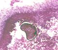
- Lesions are usually up to 2mm in diameter and contain asteroid bodies with a germinated spore in the centre
- Acute infection causes miliary lesions
- Chronic infections causes granulomatous and calcified lesions
- Horses:
- Guttural pouch mycosis common
- Infection can cause abortion
- May cause COPD
- Dogs, cats and sheep:
- Infections occur, but infrequently
- lungs and nasal cavity most usually affected
- Disseminated form with granulomas and infarcts can occur in dogs
- Pulmonary and intersitital forms can occur in cats
- Humans:
- Primary and secondary infections
- lungs, skin, nasal sinuses, external ear, bronchi, bones and meninges all affected
- Infection occurs most frequently in immunocompromised patients
- Grows on Sabauraud's Dextrose and Blood agar
- White colonies intitially which turn green, then dark green, flat and velvety
- Colony colour varies with species
- Also grows on Czapek-Dox agar and 2% malt extract agar supplemented with antibacterial antibiotics
- Microscopically:
- Conidiophores with large terminal vesicles (only visible in the lungs and air sacs where there is access to oxygen)
- Vesicle shape varies depending on the species
- Is a common contaminant so repeated tests should be done for a definitive diagnosis
- Conidiophores with large terminal vesicles (only visible in the lungs and air sacs where there is access to oxygen)
- Serology:
- Gel immunodiffusion for canine nasal asper
- Treatment:
- Surgery
- Antifungal drugs
- Pathology:
- Aspergillus fumigatus causes rhinitis, respiratory tract inflammation and sinusitis
- Sometimes appears on lesions of ethmoidal haematoma


