Difference between revisions of "Pancreatitis"
Fiorecastro (talk | contribs) |
|||
| (21 intermediate revisions by 2 users not shown) | |||
| Line 1: | Line 1: | ||
| − | {{ | + | {{OpenPagesTop}} |
| − | |||
==Introduction== | ==Introduction== | ||
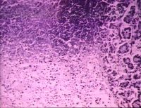
| + | [[Image:Pancreatitis.jpg|right|thumb|200px|<small><center>Pancreatitis (Image sourced from Bristol Biomed Image Archive with permission)</center></small>]] | ||
Pancreatitis occurs following activation of digestive enzymes within the [[Pancreas - Anatomy & Physiology|pancreas]] leading to autodigestion of the gland. Can be referred to as acute or chronic pancreatitis. | Pancreatitis occurs following activation of digestive enzymes within the [[Pancreas - Anatomy & Physiology|pancreas]] leading to autodigestion of the gland. Can be referred to as acute or chronic pancreatitis. | ||
| Line 22: | Line 22: | ||
Cats mainly suffer from mild chronic interstitial pancreatitis. | Cats mainly suffer from mild chronic interstitial pancreatitis. | ||
| − | |||
| − | |||
| − | + | == Acute Haemorrhagic Pancreatitis == | |
| + | |||
| + | This term is often interchangeable with [[Pancreatic Necrosis, Acute|acute pancreatic necrosis]] or '''acute pancreatitis'''. The condition can be mild or severe, non-fatal or fatal. It usually occurs as a sudden onset condition, often after ingestion of a meal rich in fat, but this depends on what species the condition occurs. | ||
| − | + | The [[Pancreas - Anatomy & Physiology#Endocrine|Islets of Langerhans]] may become involved thus causing the signs if insulin insufficiency. Pancreatitis may be initiated by trauma which initiates the leakage of enzymes. It can also present as recurrent acute pancreatitis - repeated inflammation with minimal permanent pathology. In the disease process, proteolytic degradation of pancreatic parenchyma, vascular damage and haemorrhage occur as well as necrosis of fat by lipolytic enzymes in the pancreas and surrounding omentum. These changes are concentrated at the periphery of lobules and infiltration by leukocytes indicates inflammation. In mild cases oedema of the interstitial tissue occurs. In more severe cases the [[Pancreas - Anatomy & Physiology|pancreas]] is haemorrhagic and oedematous with greyish white areas of necrosis and this may be interspersed with normal parenchyma. The [[Peritoneal Cavity - Anatomy & Physiology|peritoneal cavity]] may contain blood-stained fluid sometimes with droplets of fat. Due to these large amounts of necrotic debris, infection by microorganisms from the [[Alimentary System Overview - Anatomy & Physiology|GIT]] is likely, causing abscesses. | |
| − | == | + | === Cats and Dogs=== |
| − | |||
| − | + | See [[Pancreatitis - Cat]] and [[Pancreatitis - Dog]] | |
| − | + | === Other Animals === | |
| − | |||
| − | ''' | + | In '''horses''', necrosis and inflammation results due to migration of parasites, usually strongyle larvae, releasing pancreatic enzymes causing autodigestion. Destructive granulomatous pancreatitis is a part of multisystemic eosinophilic epitheliotrophic syndrome. |
| − | + | In '''pigs''' suppuration of the pancreas can occasionally arise as an extension from nearby infection, eg. peritonitis and perforated oesophageal ulcers. | |
| − | + | == Chronic Interstitial Pancreatitis == | |
| − | + | Chronic pancreatitis often occurs following ongoing inflammation with progression to irreversible damage and impaired function. There is usually fibrosis and reduction in acinar mass. This condition can occur in all species as a consequence of obstruction of the pancreatic ducts, [[Vitamin A Deficiency|vitamin A deficiency]] may predispose to this. The condition is most common in the dog, but also in cat, horse and cattle. The [[Pancreas - Anatomy & Physiology#Endocrine|islets of Langerhans]] tend to be preserved. If chronic pancreatitis persisits it can lead to [[Exocrine Pancreatic Insufficiency]] (EPI). In cats, chronic pancreatitis can also lead to [[Diabetes Mellitus]] developing. | |
| − | |||
| − | + | === Cats and Dogs=== | |
| − | |||
| − | |||
| − | + | See [[Pancreatitis - Cat]] and [[Pancreatitis - Dog]] | |
| − | === | + | === Other Animals === |
| − | |||
| − | '''In | + | '''In sheep''' |
| − | + | Necrosis of [[Pancreas - Anatomy & Physiology#Exocrine|exocrine pancreatic cells]] followed by fibrosis can be caused by zinc toxicosis. Focal pancreatitis may occur during [[Foot_and_Mouth_Disease|Foot and Mouth disease]] resulting in [[DM|diabetes mellitus]] during recovery. | |
| − | + | '''In horses''' | |
| − | ''' | ||
| − | |||
| − | |||
| − | + | Chronic pancreatitis can occur sporadically and is usually a consequence of [[Pancreas - Parasitic Pathology|parasitic migration]] or from ascending bacterial infection of pancreatic ducts. It can occur alongside '''chronic eosinophilic gastroenteritis''' and is usually clinically silent. Organ tends to be replaced by scar tissue. | |
| − | ''' | + | '''In cattle''' |
| − | |||
| − | + | Focal pancreatitis may occur during [[Foot_and_Mouth_Disease|Foot and Mouth disease]] resulting in [[DM|diabetes mellitus]] during recovery.<br>{{Learning | |
| − | + | |Vetstream = [https://www.vetstream.com/felis/search?s=pancreatitis Pancreatitis] | |
| − | + | |literature search = [http://www.cabdirect.org/search.html?q=title%3A%28%22pancreatitis%22%29+AND+%28od%3A%28cats%29+OR+title%3A%28dogs%29%29&fq=sc%3A%22ve%22 Pancreatitis in cats and dogs publications] | |
| + | }} | ||
| − | + | {{Chapter}} | |
| + | {{Mansonchapter | ||
| + | |chapterlink = http://www.mansonpublishing.co.uk/book-images/9781840761115_sample.pdf | ||
| + | |chaptername = Acute Pancreatitis | ||
| + | |book = Clinical Medicine of the Dog and Cat, 2nd edition | ||
| + | |author = Michael Schaer | ||
| + | |isbn = 9781840761115 | ||
| + | }} | ||
| − | == | + | ==References== |
| − | |||
| − | |||
| − | + | Andrews, A.H, Blowey, R.W, Boyd, H and Eddy, R.G. (2004) '''Bovine Medicine''' (Second edition), ''Blackwell Publishing'' | |
| − | + | Bertone, J. (2006) '''Equine Geriatric Medicine and Surgery''', ''Elsevier'' | |
| − | + | Blood, D.C. and Studdert, V. P. (1999) '''Saunders Comprehensive Veterinary Dictionary''' (2nd Edition), ''Elsevier Science'' | |
| − | + | Brown, C.M, Bertone, J.J. (2002) '''The 5-Minute Veterinary Consult- Equine''', Lippincott, ''Williams & Wilkins'' | |
| − | |||
| − | + | Cowart, R.P. and Casteel, S.W. (2001) '''An Outline of Swine diseases: a handbook,''' ''Wiley-Blackwell'' | |
| − | + | Ettinger, S.J. and Feldman, E. C. (2000) '''Textbook of Veterinary Internal Medicine Diseases of the Dog and Cat''' Volume 2 (Fifth Edition), ''W.B. Saunders Company'' | |
| − | + | Ettinger, S.J, Feldman, E.C. (2005) '''Textbook of Veterinary Internal Medicine''' (6th edition, volume 2), ''W.B. Saunders Company'' | |
| − | |||
| − | + | Fossum, T. W. et. al. (2007) '''Small Animal Surgery''' (Third Edition), ''Mosby Elsevier'' | |
| − | |||
| − | + | Hall, E.J, Simpson, J.W. and Williams, D.A. (2005) '''BSAVA Manual of Canine and Feline Gastroenterology (2nd Edition),''' ''BSAVA'' | |
| − | |||
| + | Jackson, G.G. and Cockcroft, P.D. (2007) '''Handbook of Pig Medicine,''' ''Saunders Elsevier'' | ||
| − | + | Knottenbelt, D.C. '''A Handbook of Equine Medicine for Final Year Students University of Liverpool''' | |
| − | |||
| − | |||
| − | + | Merck & Co (2008) '''The Merck Veterinary Manual''' ''Merial'' | |
| − | + | ||
| + | Nelson, R.W. and Couto, C.G. (2009) '''Small Animal Internal Medicine''' (Fourth Edition) ''Mosby Elsevier'' | ||
| + | |||
| + | Sturgess, K. (2003) '''Notes on Feline Internal Medicine''' ''Blackwell Publishing'' | ||
| + | |||
| + | Tilley, L.P. and Smith, F.W.K.(2004) '''The 5-minute Veterinary Consult''' (Third edition) Lippincott, ''Williams & Wilkins'' | ||
| + | |||
| + | |||
| + | {{review}} | ||
| + | |||
| + | {{OpenPages}} | ||
| − | + | [[Category:Pancreas_-_Inflammatory_Pathology]][[Category:Pancreatic Diseases - Dog]][[Category:Pancreatic Diseases - Cat]] | |
| − | |||
| − | + | [[Category:Pancreatic_Diseases_-_Pig]] [[Category:To_Do_-_Review]] [[Category:Pancreatic_Diseases_-_Horse]][[Category:Pancreatic_Diseases_-_Sheep]] | |
| − | [[Category: | ||
| − | [[Category: | ||
Latest revision as of 14:15, 16 March 2022
Introduction
Pancreatitis occurs following activation of digestive enzymes within the pancreas leading to autodigestion of the gland. Can be referred to as acute or chronic pancreatitis.
Acute pancreatitis is rapid onset inflammation of the pancreas with little or no pathological changes occurring post recovery. This may completely resolve or 'wax and wane' in the future.
Chronic pancreatitis is continued inflammation leading to irreversible pathological changes (fibrosis, atrophy) and possible decreases in function.
The specific cause is usually idiopathic but several risk factors exist including:
A Nutritional basis which refers to obesity, low protein and high fat diets, feeding of ethionine and hypertriglyceridaemia.
Drugs and toxins including L-asparginase, oestrogen, azathioprine, potassium bromide, furosemide, thiazide diuretics, salicylates, tetracyclines, sulphonamides, vinca alkaloids, zinc toxicosis, cholinesterase inhibitor insecticides, cholinergic agonist and hypercalcaemia.
Pancreatic duct obstruction which is caused by biliary calculi, sphincter spasm, duct wall oedema, duodenal wall oedema, neoplasia, parasites, trauma and iatrogenic reasons.
Duodenal juice reflux, pancreatic trauma, ischaemia and reperfusion which includes duodenal juice reflux into the pancreatic duct, surgical intervention, shock, anaemia, venous occlusion and hypotension.
Other risk factors include parasitic (babesiosis), viral, mycoplasmal, end stage renal disease, liver disease and auto-immune diseases.
Cats mainly suffer from mild chronic interstitial pancreatitis.
Acute Haemorrhagic Pancreatitis
This term is often interchangeable with acute pancreatic necrosis or acute pancreatitis. The condition can be mild or severe, non-fatal or fatal. It usually occurs as a sudden onset condition, often after ingestion of a meal rich in fat, but this depends on what species the condition occurs.
The Islets of Langerhans may become involved thus causing the signs if insulin insufficiency. Pancreatitis may be initiated by trauma which initiates the leakage of enzymes. It can also present as recurrent acute pancreatitis - repeated inflammation with minimal permanent pathology. In the disease process, proteolytic degradation of pancreatic parenchyma, vascular damage and haemorrhage occur as well as necrosis of fat by lipolytic enzymes in the pancreas and surrounding omentum. These changes are concentrated at the periphery of lobules and infiltration by leukocytes indicates inflammation. In mild cases oedema of the interstitial tissue occurs. In more severe cases the pancreas is haemorrhagic and oedematous with greyish white areas of necrosis and this may be interspersed with normal parenchyma. The peritoneal cavity may contain blood-stained fluid sometimes with droplets of fat. Due to these large amounts of necrotic debris, infection by microorganisms from the GIT is likely, causing abscesses.
Cats and Dogs
See Pancreatitis - Cat and Pancreatitis - Dog
Other Animals
In horses, necrosis and inflammation results due to migration of parasites, usually strongyle larvae, releasing pancreatic enzymes causing autodigestion. Destructive granulomatous pancreatitis is a part of multisystemic eosinophilic epitheliotrophic syndrome.
In pigs suppuration of the pancreas can occasionally arise as an extension from nearby infection, eg. peritonitis and perforated oesophageal ulcers.
Chronic Interstitial Pancreatitis
Chronic pancreatitis often occurs following ongoing inflammation with progression to irreversible damage and impaired function. There is usually fibrosis and reduction in acinar mass. This condition can occur in all species as a consequence of obstruction of the pancreatic ducts, vitamin A deficiency may predispose to this. The condition is most common in the dog, but also in cat, horse and cattle. The islets of Langerhans tend to be preserved. If chronic pancreatitis persisits it can lead to Exocrine Pancreatic Insufficiency (EPI). In cats, chronic pancreatitis can also lead to Diabetes Mellitus developing.
Cats and Dogs
See Pancreatitis - Cat and Pancreatitis - Dog
Other Animals
In sheep
Necrosis of exocrine pancreatic cells followed by fibrosis can be caused by zinc toxicosis. Focal pancreatitis may occur during Foot and Mouth disease resulting in diabetes mellitus during recovery.
In horses
Chronic pancreatitis can occur sporadically and is usually a consequence of parasitic migration or from ascending bacterial infection of pancreatic ducts. It can occur alongside chronic eosinophilic gastroenteritis and is usually clinically silent. Organ tends to be replaced by scar tissue.
In cattle
Focal pancreatitis may occur during Foot and Mouth disease resulting in diabetes mellitus during recovery.
| Pancreatitis Learning Resources | |
|---|---|
To reach the Vetstream content, please select |
Canis, Felis, Lapis or Equis |
 Search for recent publications via CAB Abstract (CABI log in required) |
Pancreatitis in cats and dogs publications |
| Sample Book Chapters | ||||
|---|---|---|---|---|
|
|
|
|
|
|
|
|
|
References
Andrews, A.H, Blowey, R.W, Boyd, H and Eddy, R.G. (2004) Bovine Medicine (Second edition), Blackwell Publishing
Bertone, J. (2006) Equine Geriatric Medicine and Surgery, Elsevier
Blood, D.C. and Studdert, V. P. (1999) Saunders Comprehensive Veterinary Dictionary (2nd Edition), Elsevier Science
Brown, C.M, Bertone, J.J. (2002) The 5-Minute Veterinary Consult- Equine, Lippincott, Williams & Wilkins
Cowart, R.P. and Casteel, S.W. (2001) An Outline of Swine diseases: a handbook, Wiley-Blackwell
Ettinger, S.J. and Feldman, E. C. (2000) Textbook of Veterinary Internal Medicine Diseases of the Dog and Cat Volume 2 (Fifth Edition), W.B. Saunders Company
Ettinger, S.J, Feldman, E.C. (2005) Textbook of Veterinary Internal Medicine (6th edition, volume 2), W.B. Saunders Company
Fossum, T. W. et. al. (2007) Small Animal Surgery (Third Edition), Mosby Elsevier
Hall, E.J, Simpson, J.W. and Williams, D.A. (2005) BSAVA Manual of Canine and Feline Gastroenterology (2nd Edition), BSAVA
Jackson, G.G. and Cockcroft, P.D. (2007) Handbook of Pig Medicine, Saunders Elsevier
Knottenbelt, D.C. A Handbook of Equine Medicine for Final Year Students University of Liverpool
Merck & Co (2008) The Merck Veterinary Manual Merial
Nelson, R.W. and Couto, C.G. (2009) Small Animal Internal Medicine (Fourth Edition) Mosby Elsevier
Sturgess, K. (2003) Notes on Feline Internal Medicine Blackwell Publishing
Tilley, L.P. and Smith, F.W.K.(2004) The 5-minute Veterinary Consult (Third edition) Lippincott, Williams & Wilkins
| This article has been peer reviewed but is awaiting expert review. If you would like to help with this, please see more information about expert reviewing. |
Error in widget FBRecommend: unable to write file /var/www/wikivet.net/extensions/Widgets/compiled_templates/wrt663367ec4cfd13_31505729 Error in widget google+: unable to write file /var/www/wikivet.net/extensions/Widgets/compiled_templates/wrt663367ec505cd5_04096798 Error in widget TwitterTweet: unable to write file /var/www/wikivet.net/extensions/Widgets/compiled_templates/wrt663367ec537128_75180908
|
| WikiVet® Introduction - Help WikiVet - Report a Problem |
