Difference between revisions of "Snake Digestive System"
Danimonster (talk | contribs) |
|||
| (4 intermediate revisions by 2 users not shown) | |||
| Line 1: | Line 1: | ||
| − | {{ | + | {{OpenPagesTop}} |
| − | |||
The digestive system consists of the oral cavity, oesophagus, stomach, small intestine, caecum (some species), colon and cloaca. | The digestive system consists of the oral cavity, oesophagus, stomach, small intestine, caecum (some species), colon and cloaca. | ||
==Oral cavity== | ==Oral cavity== | ||
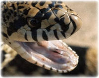
[[Image:Open_mouth.jpg|200px|thumb|right|'''Sheathed tongue of a bullsnake''' - (Copyright © RVC)]] | [[Image:Open_mouth.jpg|200px|thumb|right|'''Sheathed tongue of a bullsnake''' - (Copyright © RVC)]] | ||
| − | The mouth of a snake can open widely by the independent movement of the jaws to accommodate relatively [[Snake Feeding|large prey]]. The mucous salivary glands moisten the mouth, lubricate prey, aid digestion and excrete salt. Venom glands that produce toxins to kill prey are modified salivary glands. The sheathed tongue lies in a diverticulum on the floor of the mouth ventral to the glottis. It functions for [[Snake Special Senses|vomeronasal chemoreception]] and plays no role in swallowing, during which is is retracted into the sheath. Snakes that lose their tongues may not feed. The surface epithelial layer or the snake's tongue is periodically [[Snake Shedding|shed]] intact and is often seen in the water bowl. The oral mucosa of snakes is paler than that of mammals since the PCV may be about half to two thirds that of mammals. | + | The mouth of a snake can open widely by the independent movement of the jaws to accommodate relatively [[Snake Feeding and Digestion|large prey]]. The mucous salivary glands moisten the mouth, lubricate prey, aid digestion and excrete salt. Venom glands that produce toxins to kill prey are modified salivary glands. The sheathed tongue lies in a diverticulum on the floor of the mouth ventral to the glottis. It functions for [[Snake Special Senses|vomeronasal chemoreception]] and plays no role in swallowing, during which is is retracted into the sheath. Snakes that lose their tongues may not feed. The surface epithelial layer or the snake's tongue is periodically [[Snake Shedding|shed]] intact and is often seen in the water bowl. The oral mucosa of snakes is paler than that of mammals since the PCV may be about half to two thirds that of mammals. |
==Oesophagus, stomach and intestines== | ==Oesophagus, stomach and intestines== | ||
| − | The oesophagus is distensible to accommodate relatively large prey (for a list of diets, see [[Snake Diet|snake | + | The oesophagus is distensible to accommodate relatively large prey (for a list of diets, see [[Snake Diet|snake diet]]). The cranial oesophagus is thinly muscled. At the level of the [[Snake Cardiovascular System|heart]], the oesophagus passes through the vascular ring formed by the left and right aortae. It possesses longitudinal folds and is covered with a columnar ciliated epithelium. The oesophagus terminates as the [[Snake Cardiovascular System|cardiac sphincter]]. The thick-walled, spindle-shaped stomach is muscular and distensible. The small intestine is relatively uncoiled but has several short transverse loops tightly enveloped by dorsal mesentery. It empties into the colon that may store faeces. The large intestine is relatively wide and is separated from the cloaca by a distinct fold. A small caecum projects from the proximal colon in Boidae. |
| − | * For information on snake digestion, [[Snake Digestion| | + | * For more information on snake digestion, see [[Snake Feeding and Digestion|snake digestion]]. |
| − | * Learn about excretion [[Lizard and Snake Excretion|here]]. | + | * Learn about snake excretion [[Lizard and Snake Excretion|here]]. |
| + | |||
==Liver, pancreas and gall bladder== | ==Liver, pancreas and gall bladder== | ||
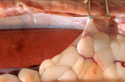
[[Image:Bumese_python_liver_+_seosa-sheath_+_fat_tissue2.jpg|180px|thumb|right|'''Liver and fat body of a [[Burmese Python|Burmese python]]''' - (Copyright © RVC)]] | [[Image:Bumese_python_liver_+_seosa-sheath_+_fat_tissue2.jpg|180px|thumb|right|'''Liver and fat body of a [[Burmese Python|Burmese python]]''' - (Copyright © RVC)]] | ||
| Line 16: | Line 16: | ||
==Cloaca== | ==Cloaca== | ||
The cloaca is divided into three parts (cranial to caudal): coprodeum, urodeum and proctodeum. The coprodeum collects faeces from the colon. The urodeum, which is the midsection of the cloaca, collects urinary waste and products of [[Snake Reproductive System|reproduction]]. The urogenital papilla is situated dorsally behind a small fold. The proctodeum is the reservoir for faecal and urinary waste before [[Lizard and Snake Excretion|excretion]] and contains the openings of the cloacal scent glands. | The cloaca is divided into three parts (cranial to caudal): coprodeum, urodeum and proctodeum. The coprodeum collects faeces from the colon. The urodeum, which is the midsection of the cloaca, collects urinary waste and products of [[Snake Reproductive System|reproduction]]. The urogenital papilla is situated dorsally behind a small fold. The proctodeum is the reservoir for faecal and urinary waste before [[Lizard and Snake Excretion|excretion]] and contains the openings of the cloacal scent glands. | ||
| + | |||
| + | {{review}} | ||
| + | |||
| + | {{OpenPages}} | ||
| + | |||
| + | |||
[[Category:Snake_Anatomy]] | [[Category:Snake_Anatomy]] | ||
Latest revision as of 16:24, 10 January 2013
The digestive system consists of the oral cavity, oesophagus, stomach, small intestine, caecum (some species), colon and cloaca.
Oral cavity
The mouth of a snake can open widely by the independent movement of the jaws to accommodate relatively large prey. The mucous salivary glands moisten the mouth, lubricate prey, aid digestion and excrete salt. Venom glands that produce toxins to kill prey are modified salivary glands. The sheathed tongue lies in a diverticulum on the floor of the mouth ventral to the glottis. It functions for vomeronasal chemoreception and plays no role in swallowing, during which is is retracted into the sheath. Snakes that lose their tongues may not feed. The surface epithelial layer or the snake's tongue is periodically shed intact and is often seen in the water bowl. The oral mucosa of snakes is paler than that of mammals since the PCV may be about half to two thirds that of mammals.
Oesophagus, stomach and intestines
The oesophagus is distensible to accommodate relatively large prey (for a list of diets, see snake diet). The cranial oesophagus is thinly muscled. At the level of the heart, the oesophagus passes through the vascular ring formed by the left and right aortae. It possesses longitudinal folds and is covered with a columnar ciliated epithelium. The oesophagus terminates as the cardiac sphincter. The thick-walled, spindle-shaped stomach is muscular and distensible. The small intestine is relatively uncoiled but has several short transverse loops tightly enveloped by dorsal mesentery. It empties into the colon that may store faeces. The large intestine is relatively wide and is separated from the cloaca by a distinct fold. A small caecum projects from the proximal colon in Boidae.
- For more information on snake digestion, see snake digestion.
- Learn about snake excretion here.
Liver, pancreas and gall bladder

The liver is elongated and is divided into several separate lobes. A characteristic of snakes is that there is a relatively long distance between the caudal tip of the liver and the gall bladder. Snakes have a well-developed gall bladder adjacent to the duodenum and caudal to the liver. Multiple bile ducts pass from the gall bladder, through the pancreas, into the duodenum. In most species, the gall bladder, pancreas and spleen are closely associated. In some snakes the pancreas may be fused with the spleen forming the splenopancreas.
Cloaca
The cloaca is divided into three parts (cranial to caudal): coprodeum, urodeum and proctodeum. The coprodeum collects faeces from the colon. The urodeum, which is the midsection of the cloaca, collects urinary waste and products of reproduction. The urogenital papilla is situated dorsally behind a small fold. The proctodeum is the reservoir for faecal and urinary waste before excretion and contains the openings of the cloacal scent glands.
| This article has been peer reviewed but is awaiting expert review. If you would like to help with this, please see more information about expert reviewing. |
Error in widget FBRecommend: unable to write file /var/www/wikivet.net/extensions/Widgets/compiled_templates/wrt693b2a9d77a673_82883373 Error in widget google+: unable to write file /var/www/wikivet.net/extensions/Widgets/compiled_templates/wrt693b2a9d7d6aa0_42494536 Error in widget TwitterTweet: unable to write file /var/www/wikivet.net/extensions/Widgets/compiled_templates/wrt693b2a9d82e932_37298551
|
| WikiVet® Introduction - Help WikiVet - Report a Problem |
