Difference between revisions of "Systemic Mycoses"
Jump to navigation
Jump to search
(Redirected page to Category:Systemic Mycoses) |
|||
| (18 intermediate revisions by 3 users not shown) | |||
| Line 1: | Line 1: | ||
| − | # | + | {{unfinished}} |
| + | |||
| + | {{toplink | ||
| + | |backcolour = | ||
| + | |linkpage =Fungi | ||
| + | |linktext =FUNGI | ||
| + | |pagetype=Bugs | ||
| + | }} | ||
| + | <br> | ||
| + | |||
| + | ==Adiaspiromycosis== | ||
| + | |||
| + | *Haplomycosis | ||
| + | |||
| + | *''Emmonsia crescens'' | ||
| + | **Does not proliferate within the animal body | ||
| + | **Each spore develops into a thick-walled spherule called an '''adiaspore''' | ||
| + | |||
| + | *''Chrysosporium parvum, C. crescens'' | ||
| + | |||
| + | *Non-contageous, pulmonary mycosis | ||
| + | |||
| + | *Worldwide | ||
| + | |||
| + | *Found in soil | ||
| + | |||
| + | *Affects burrowing rodents and small animals | ||
| + | |||
| + | *Respiratory infection | ||
| + | |||
| + | *Spetate hyphae with large numbers of small, round conidia either singly or in groups on the ends of the short conidiospores can be seen | ||
| + | |||
| + | *Dimorphic | ||
| + | |||
| + | *Grows on Sabauraud's Dextrose agar and Blood agar | ||
| + | ==Aspergillosis== | ||
| + | [[Image:Aspergillus cleistothecia.jpg|thumb|right|150px|Aspergillus cleistothecia - Copyright Professor Andrew N. Rycroft, BSc, PHD, C. Biol.F.I.Biol., FRCPath]] | ||
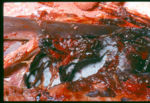
| + | [[Image:Aspergillus swan.jpg|thumb|right|150px|Aspergillus in a swan - Copyright Professor Andrew N. Rycroft, BSc, PHD, C. Biol.F.I.Biol., FRCPath]] | ||
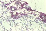
| + | [[Image:Aspergillus in vivo.jpg|thumb|right|150px|Aspergillus in vivo - Copyright Professor Andrew N. Rycroft, BSc, PHD, C. Biol.F.I.Biol., FRCPath]] | ||
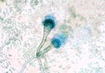
| + | [[Image:Aspergillus sporing heads.jpg|thumb|right|150px|Aspergillus sporing heads - Copyright Professor Andrew N. Rycroft, BSc, PHD, C. Biol.F.I.Biol., FRCPath]] | ||
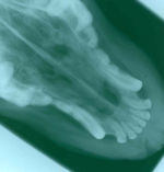
| + | [[Image:Canine nasal asper radiograph.jpg|thumb|right|150px|Canine nasal aspergillus radiograph - Copyright Professor Andrew N. Rycroft, BSc, PHD, C. Biol.F.I.Biol., FRCPath]] | ||
| + | *Worldwide | ||
| + | |||
| + | *Widely found in nature | ||
| + | **Colonise a wide range of substrates under different environmental conditions | ||
| + | **Abundant in hay, straw and grain which have heated during storage | ||
| + | |||
| + | *Common laboratory contaminants | ||
| + | |||
| + | *Pathogenic species include ''Aspergillus fumigatus, A. flavus, A. nidulans, A.niger'' and ''A. terreus'' | ||
| + | |||
| + | *May cause primary or secondary disease | ||
| + | **Infection may be acute, chronic or benign | ||
| + | |||
| + | *Avians: | ||
| + | **Diffuse infection of the [[Avian Respiration - Anatomy & Physiology#Air Sacs|air sacs]] | ||
| + | **Diffuse pneumonic form | ||
| + | **Nodular form involving the [[Avian Respiration - Anatomy & Physiology#Avian Lungs|lungs]] | ||
| + | **Spores are inhaled | ||
| + | **Yellow nodules in the [[Avian Respiration - Anatomy & Physiology#Avian Lungs|lungs]] and [[Avian Respiration - Anatomy & Physiology#Air Sacs|air sacs]] | ||
| + | **The acute form usually affects young birds and is rapidly fatal (within 24-48 hours) | ||
| + | ***Signs include [[Intestine Diarrhoea - Pathology|diarrhoea]], listlessness, pyrexia, loss of appetite and loss of condition | ||
| + | ***Sometimes convulsions may occur | ||
| + | ***Resembles Pullorum disease | ||
| + | **The chronic form usually occurs in adult birds and is sporadic, presenting with milder clinical signs | ||
| + | |||
| + | *Cattle: | ||
| + | **Infection can cause abortion and ocular infections | ||
| + | **Infections involve the [[Female Reproductive Tract -The Uterus - Anatomy & Physiology|uterus]], [[Foetal Membranes - Anatomy & Physiology|fetal membranes]] and fetal skin | ||
| + | **Lesions are usually up to 2mm in diameter and contain asteroid bodies with a germinated spore in the centre | ||
| + | ***Acute infection causes miliary lesions | ||
| + | ***Chronic infections causes granulomatous and calcified lesions | ||
| + | |||
| + | *Horses: | ||
| + | **[[Guttural Pouches Inflammatory - Pathology|Guttural pouch mycosis]] common | ||
| + | **Infection can cause abortion | ||
| + | **May cause [[Bronchi and Bronchioles Inflammatory - Pathology#Chronic obstructive pulmonary disease (COPD)|COPD]] | ||
| + | |||
| + | *Dogs, cats and sheep: | ||
| + | **Infections occur, but infrequently | ||
| + | **[[Lungs - Anatomy & Physiology|lungs]] and [[Nasal cavity - Anatomy & Physiology|nasal cavity]] most usually affected | ||
| + | **Disseminated form with granulomas and infarcts can occur in dogs | ||
| + | **Pulmonary and intersitital forms can occur in cats | ||
| + | |||
| + | *Humans: | ||
| + | **Primary and secondary infections | ||
| + | **[[Lungs - Anatomy & Physiology|lungs]], [[Skin - Anatomy & Physiology|skin]], [[Nasal cavity - Anatomy & Physiology|nasal sinuses]], [[Special Senses - Auditory - Anatomy & Physiology#Outer Ear|external ear]], [[Bronchi and bronchioles - Anatomy & Physiology|bronchi]], [[Bones and Cartilage - Anatomy & Physiology|bones]] and meninges all affected | ||
| + | **Infection occurs most frequently in immunocompromised patients | ||
| + | |||
| + | *Grows on Sabauraud's Dextrose and Blood agar | ||
| + | **White colonies intitially which turn green, then dark green, flat and velvety | ||
| + | **Colony colour varies with species | ||
| + | |||
| + | *Also grows on Czapek-Dox agar and 2% malt extract agar supplemented with antibacterial antibiotics | ||
| + | |||
| + | *Microscopically: | ||
| + | **Conidiophores with large terminal vesicles (only visible in the [[Lungs - Anatomy & Physiology|lungs]] and air sacs where there is access to oxygen) | ||
| + | ***Vesicle shape varies depending on the species | ||
| + | **Is a common contaminant so repeated tests should be done for a definitive diagnosis | ||
| + | |||
| + | *Serology: | ||
| + | **Gel immunodiffusion for canine nasal asper | ||
| + | |||
| + | *Treatment: | ||
| + | **Surgery | ||
| + | **Antifungal drugs | ||
| + | ***[[Antifungal Drugs#The Azoles|Ketoconazole]], [[Antifungal Drugs#Polyene Antifungals|Nystatin]], [[Antifungal Drugs#Polyene Antifungals|Amphotericin B]], [[Antifungal Drugs#Flucytosine|5-fluorocytosine]], [[Antifungal Drugs#The Azoles|Thiabendazole]] | ||
| + | |||
| + | *Pathology: | ||
| + | **''Aspergillus fumigatus'' causes [[Nasal Cavity Inflammatory - Pathology#Infectious causes of rhinitis|rhinitis]], [[Respiratory Fungal Infections - Pathology#|respiratory tract inflammation]] and [[Paranasal Sinuses Inflammatory - Pathology#Infectious causes of sinusitis|sinusitis]] | ||
| + | **Sometimes appears on [[Nasal Cavity Hyperplastic and Neoplastic - Pathology#Progressive ethmoidal haematoma|lesions of ethmoidal haematoma]] | ||
| + | |||
| + | ==Blastomycosis== | ||
| + | |||
| + | *North America | ||
| + | **Most common in the North-Central and South-Eastern states | ||
| + | |||
| + | *Caused by ''Blastomyces dermatitidis'' | ||
| + | |||
| + | *Widespread in soil | ||
| + | |||
| + | *Respiratory infection | ||
| + | |||
| + | *Lesions start in the [[Lungs - Anatomy & Physiology|lungs]] | ||
| + | **Haematogenous dissemination | ||
| + | **Can be found in lesions in the eyes, brain, bones and genitalia | ||
| + | **Fatal if not treated | ||
| + | |||
| + | *Lesions are also found on the skin | ||
| + | *These may ulcerate | ||
| + | |||
| + | *Granulomatous nodules | ||
| + | |||
| + | *Affects mainly dogs (and humans) | ||
| + | **Can affect cats, horses, dolphins, ferrets and sealions but is rare in these species | ||
| + | |||
| + | *Microscopically: | ||
| + | **Large, spherical, thick-walled cells | ||
| + | **Single buds connected to a mother cell by a wide base | ||
| + | **Double contoured effect of cells | ||
| + | |||
| + | *Grows on Sabauraud's Dextrose and Blood agar | ||
| + | **On Sabauraud's Dextrose colonies appear moist and grey with a white cotton-like mycelium which turns tan, brown and then black | ||
| + | ***Septate hyphae | ||
| + | ***Small, oval/pyriform conidia | ||
| + | ***Older cultures have thickened walls | ||
| + | **On Blood agar colonies are creamy in colour, waxy and wrinkled | ||
| + | ***Thick-walled budding yeast cells can be seen | ||
| + | |||
| + | *Diagnosis: | ||
| + | **Complement fixation test | ||
| + | **Falling antibody titres indicate a poor prognosis | ||
| + | **ELISA and counterimmunoelectrophoresis can also be used | ||
| + | |||
| + | *Treatment: | ||
| + | **[[Antifungal Drugs#Polyene Antifungals|Amphotericin B]] | ||
| + | **[[Antifungal Drugs#The Azoles|Imidazoles]] | ||
| + | |||
| + | ==Coccidioidomycosis== | ||
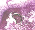
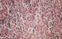
| + | [[Image:Coccidioidomycosis.jpg|thumb|right|200px|Coccidioidomycosis spherule histopathology - Copyright Professor Andrew N. Rycroft, BSc, PHD, C. Biol.F.I.Biol., FRCPath]] | ||
| + | *''Coccidioides immitis'' | ||
| + | |||
| + | *Ocurs in the soil | ||
| + | **Respiratory infections | ||
| + | **Most commonly seen following dust storms | ||
| + | |||
| + | *Occurs in arid regions | ||
| + | **E.g. South West USA and Mexico | ||
| + | |||
| + | *Non-contageous, systemic mycosis | ||
| + | |||
| + | *Affects dogs, cattle, sheep and humans | ||
| + | |||
| + | *Mainly affects the [[Lungs - Anatomy & Physiology|lungs]] | ||
| + | **Dissemination can occur to other organs | ||
| + | |||
| + | *Causes nodule or granuloma formation | ||
| + | **Localised | ||
| + | **Gross lesions resemble [[Mycobacteria spp.#Bovine tuberculosis|Tb]] in cattle as are usually seen in the bronchial and mediastinal [[Lymph Nodes - Anatomy & Physiology|lymph nodes]] and occasionally [[Lungs - Anatomy & Physiology|lungs]] | ||
| + | **Dissemination can occur, especially in primates and dogs, to the [[Lungs - Anatomy & Physiology|lungs]], [[Liver - Anatomy & Physiology|liver]], [[Spleen - Anatomy & Physiology|spleen]], [[Nervous System - CNS - Anatomy & Physiology|brain]] and [[Bones and Cartilage - Anatomy & Physiology|bones]] | ||
| + | |||
| + | *Thick-walled spherules in tissue | ||
| + | **Large sporangia burst leaving 'ghost' spherules | ||
| + | |||
| + | *Saprophytic phase consists of coarse, septate, branching hyphae which fragment into thick-walled, barrel-shaped arthrospores which alternate with empty cells | ||
| + | **Stained by Lactose Phenol Cotton Blue | ||
| + | |||
| + | *Grows on Sabouraud's Dextrose agar and Blood agar | ||
| + | **Flat, moist colonies which develop a coarse, cotton-like aerial mycelium which varies from white to brown in colour | ||
| + | |||
| + | *Complement fixation test, latex agglutination and immunodiffusion tests can all be used | ||
| + | **A positive skin test indicates exposure | ||
| + | |||
| + | ==Entomophthoromycisus== | ||
| + | |||
| + | *Basidiobolmycosis | ||
| + | |||
| + | *Caused by ''Basidiobolus'' and ''Conidiobulus'' fungi | ||
| + | |||
| + | *Causes ulcerative granulomas in subcutaneous tissue | ||
| + | |||
| + | *Affects the oral and nasal mucous membranes | ||
| + | |||
| + | *''Basidiobolus'' causes large lesions which may involve skin on the head, neck and chest | ||
| + | **Fistulous tracts | ||
| + | **Extends to [[Lymph Nodes - Anatomy & Physiology|lymph nodes]] | ||
| + | |||
| + | *Produce flat, waxy colonies which become white and fizzy over time | ||
| + | |||
| + | *Microscopically: | ||
| + | **Septate hyphae | ||
| + | |||
| + | *Treatment: | ||
| + | **Surgical excision | ||
| + | **[[Antifungal Drugs#Polyene Antifungals|Amphotericin B]] or [[Antifungal Drugs#The Azoles|Ketoconazole]] | ||
| + | |||
| + | ==Histoplasmosis== | ||
| + | {| align="right" | ||
| + | |<gallery>Image:Histoplasmosis canine spleen.jpg|<center><p>'''Histoplasmosis in a canine spleen'''</p><sup>Copyright Professor Andrew N. Rycroft, BSc, PHD, C. Biol.F.I.Biol., FRCPath</sup></center> | ||
| + | Image:Histoplasmosis canine spleen silver stain.jpg|<center><p>'''Histoplasmosis in a canine spleen using silver stain'''</p><sup>Copyright Professor Andrew N. Rycroft, BSc, PHD, C. Biol.F.I.Biol., FRCPath</sup></center></gallery> | ||
| + | |} | ||
| + | |||
| + | *''Histoplasma capsulatum'' | ||
| + | |||
| + | *Non-contageous, systemic mycosis | ||
| + | |||
| + | *Commonly pulmonary infections occur | ||
| + | **Other organs can be involved | ||
| + | **Involves the reticuloendothelial system | ||
| + | **Intestinal form can also occur | ||
| + | |||
| + | *Acute and chronic disease can occur | ||
| + | |||
| + | *Endemic to the USA | ||
| + | **Isolated cases have been reported in Europe | ||
| + | |||
| + | *Respiratory infection | ||
| + | **Infection via ingestion can also occur | ||
| + | |||
| + | *Affects dogs, cats, cattle, horses and humans | ||
| + | |||
| + | *Found in soil contaminated by bird droppings, decaying vegetation and in caves inhabited by bats | ||
| + | {| align="right" | ||
| + | |<gallery>Image:Histoplasmosis lung.jpg|<center><p>'''Histoplasmosis lesions in lungs'''</p><sup>Copyright Professor Andrew N. Rycroft, BSc, PHD, C. Biol.F.I.Biol., FRCPath</sup></center> | ||
| + | Image:Histoplasmosis phagocyte.jpg|<center><p>'''Histoplasmosis phagocyte'''</p><sup>Copyright Professor Andrew N. Rycroft, BSc, PHD, C. Biol.F.I.Biol., FRCPath</sup></center></gallery> | ||
| + | |} | ||
| + | *Fine, branching, septate hyphae with smooth-walled pyriform to spherical microconidia and large, thick-walled tuberculate macroconidia on simple conidiophores | ||
| + | |||
| + | *Dimorphic fungi | ||
| + | |||
| + | *Hard to demonstrate in smears as the organisms is very small | ||
| + | **Stain with Giemsa or Wright and examine under oil immersion lens | ||
| + | |||
| + | *Present intracellularly in [[Macrophages - WikiBlood|macrophages]] as oval yeast cells with few buds | ||
| + | **Clear halo is seen around the darker staining central material | ||
| + | |||
| + | *Grows on Sabouraud's Dextrose agar | ||
| + | **Creamy white colonies, turning tan coloured and then brown | ||
| + | |||
| + | *Also grows on Blood agar | ||
| + | **Small, white yeast-like colonies | ||
| + | |||
| + | *Test using immunodiffusion, complement fixation and counterimmunoelectrophoresis | ||
| + | **Skin test of little value as it only indicates exposure | ||
| + | {| align="right" | ||
| + | |<gallery>Image:Histoplasmosis tuberculate chlamydospores.jpg|<center><p>'''Histoplasmosis tuberculate chlamydospores'''</p><sup>Copyright Professor Andrew N. Rycroft, BSc, PHD, C. Biol.F.I.Biol., FRCPath</sup></center></gallery> | ||
| + | |} | ||
| + | *Treatment with [[Antifungal Drugs#Polyene Antifungals|Amphotericin B]] | ||
| + | **If [[Antifungal Drugs#Polyene Antifungals|Amphotericin B]] is contra-indicated, [[Antifungal Drugs#Imidazoles|imidazoles]] can be given orally | ||
| + | |||
| + | *The prognosis is poor in acute and disseminated cases | ||
| + | |||
| + | ==Zygomycosis== | ||
| + | |||
| + | *Also known as mucormycosis, hyphomycosis and phycomycosis | ||
| + | |||
| + | *Caused by strains of ''Mucor, Absidia, Rhizopus'' and ''Mortierella'' | ||
| + | **''Mucor circinelloides''(rare), ''Rhizomucor pusillus'' and ''R. meihi'' | ||
| + | **''Absidia corymbifera'' often causes zygomycosis in cattle and pigs | ||
| + | **''Rhizopus arrhizus, R. microsporus'' and ''R. rhizopodormis'' | ||
| + | **''Mortierella wolfi'' implicated in bovine abortion (mycotic placentitis), ''M. hygrophila'' in fowl and ''M.polycephala'' in cattle | ||
| + | |||
| + | *Occurs widely in nature | ||
| + | |||
| + | *Infection is by inhalation and ingestion | ||
| + | |||
| + | *Infects [[Lymph Nodes - Anatomy & Physiology|lymph nodes]] of the [[Cardiorespiratory System - Anatomy & Physiology|respiratory]] and [[Alimentary - Anatomy & Physiology|alimentary tract]] | ||
| + | **[[Lymph Nodes - Anatomy & Physiology|Lymph nodes]] enlarge and become caseous | ||
| + | **Can cause [[Alimentary - Anatomy & Physiology#Stomach|stomach]] and [[Small Intestine - Anatomy & Physiology|intestinal]] ulcers | ||
| + | |||
| + | *Granulomatous lesions which can ulcerate | ||
| + | |||
| + | *Mostly localised lesions but can be generalised | ||
| + | |||
| + | *Pigs | ||
| + | **Mediastinal and submandibular [[Lymph Nodes - Anatomy & Physiology|lymph nodes]] lesions | ||
| + | **Embolic tumours in the [[Liver - Anatomy & Physiology|liver]] and [[Lungs - Anatomy & Physiology|lungs]] | ||
| + | **Can also be present in gastric ulcers | ||
| + | |||
| + | *Cattle | ||
| + | **Bronchial, mesenteric and mediastinal [[Lymph Nodes - Anatomy & Physiology|lymph nodes]] lesions | ||
| + | **Ulcers of the [[Nasal cavity - Anatomy & Physiology|nasal cavity]] and [[The Abomasum - Anatomy & Physiology|abomasum]] also occur | ||
| + | **Often contaminate the [[Gestation -Placenta - Anatomy & Physiology|placenta]] | ||
| + | |||
| + | *Horses, dogs, cats, sheep, mink, guinea-pigs and mice can also be infected | ||
| + | |||
| + | *Microscopically: | ||
| + | **Fragments of non-septate hyphae which are branched and coarse | ||
| + | **''Rhizomucor'' produce a thick, grey mycelium and have short, black, spherical sporangia | ||
| + | **''Mucor'' produce thick, colourless mycelium with no rhizoids. Globose spoangia with small spores are present and sporagiospores are simple or branched. | ||
| + | **''Absidia'' resemble ''Rhizopus'' grossly | ||
| + | **''Mortierella'' produce white, velvet colonies on Sabouraud's Dextrose and Blood agar | ||
| + | |||
| + | *Grows on Sabauraud's Dextrose agar | ||
| + | **Common contaminants | ||
| + | |||
| + | *Treatment is with [[Antifungal Drugs#Polyene Antifungals|Amphotericin B]] | ||
| + | **Surgery is also an option in treatment | ||
| + | |||
| + | ==Further Links== | ||
| + | |||
| + | *[[Antifungal Drugs]] | ||
Revision as of 19:00, 4 June 2009
| This article is still under construction. |
|
|
Adiaspiromycosis
- Haplomycosis
- Emmonsia crescens
- Does not proliferate within the animal body
- Each spore develops into a thick-walled spherule called an adiaspore
- Chrysosporium parvum, C. crescens
- Non-contageous, pulmonary mycosis
- Worldwide
- Found in soil
- Affects burrowing rodents and small animals
- Respiratory infection
- Spetate hyphae with large numbers of small, round conidia either singly or in groups on the ends of the short conidiospores can be seen
- Dimorphic
- Grows on Sabauraud's Dextrose agar and Blood agar
Aspergillosis
- Worldwide
- Widely found in nature
- Colonise a wide range of substrates under different environmental conditions
- Abundant in hay, straw and grain which have heated during storage
- Common laboratory contaminants
- Pathogenic species include Aspergillus fumigatus, A. flavus, A. nidulans, A.niger and A. terreus
- May cause primary or secondary disease
- Infection may be acute, chronic or benign
- Avians:
- Diffuse infection of the air sacs
- Diffuse pneumonic form
- Nodular form involving the lungs
- Spores are inhaled
- Yellow nodules in the lungs and air sacs
- The acute form usually affects young birds and is rapidly fatal (within 24-48 hours)
- Signs include diarrhoea, listlessness, pyrexia, loss of appetite and loss of condition
- Sometimes convulsions may occur
- Resembles Pullorum disease
- The chronic form usually occurs in adult birds and is sporadic, presenting with milder clinical signs
- Cattle:
- Infection can cause abortion and ocular infections
- Infections involve the uterus, fetal membranes and fetal skin
- Lesions are usually up to 2mm in diameter and contain asteroid bodies with a germinated spore in the centre
- Acute infection causes miliary lesions
- Chronic infections causes granulomatous and calcified lesions
- Horses:
- Guttural pouch mycosis common
- Infection can cause abortion
- May cause COPD
- Dogs, cats and sheep:
- Infections occur, but infrequently
- lungs and nasal cavity most usually affected
- Disseminated form with granulomas and infarcts can occur in dogs
- Pulmonary and intersitital forms can occur in cats
- Humans:
- Primary and secondary infections
- lungs, skin, nasal sinuses, external ear, bronchi, bones and meninges all affected
- Infection occurs most frequently in immunocompromised patients
- Grows on Sabauraud's Dextrose and Blood agar
- White colonies intitially which turn green, then dark green, flat and velvety
- Colony colour varies with species
- Also grows on Czapek-Dox agar and 2% malt extract agar supplemented with antibacterial antibiotics
- Microscopically:
- Conidiophores with large terminal vesicles (only visible in the lungs and air sacs where there is access to oxygen)
- Vesicle shape varies depending on the species
- Is a common contaminant so repeated tests should be done for a definitive diagnosis
- Conidiophores with large terminal vesicles (only visible in the lungs and air sacs where there is access to oxygen)
- Serology:
- Gel immunodiffusion for canine nasal asper
- Treatment:
- Surgery
- Antifungal drugs
- Pathology:
- Aspergillus fumigatus causes rhinitis, respiratory tract inflammation and sinusitis
- Sometimes appears on lesions of ethmoidal haematoma
Blastomycosis
- North America
- Most common in the North-Central and South-Eastern states
- Caused by Blastomyces dermatitidis
- Widespread in soil
- Respiratory infection
- Lesions start in the lungs
- Haematogenous dissemination
- Can be found in lesions in the eyes, brain, bones and genitalia
- Fatal if not treated
- Lesions are also found on the skin
- These may ulcerate
- Granulomatous nodules
- Affects mainly dogs (and humans)
- Can affect cats, horses, dolphins, ferrets and sealions but is rare in these species
- Microscopically:
- Large, spherical, thick-walled cells
- Single buds connected to a mother cell by a wide base
- Double contoured effect of cells
- Grows on Sabauraud's Dextrose and Blood agar
- On Sabauraud's Dextrose colonies appear moist and grey with a white cotton-like mycelium which turns tan, brown and then black
- Septate hyphae
- Small, oval/pyriform conidia
- Older cultures have thickened walls
- On Blood agar colonies are creamy in colour, waxy and wrinkled
- Thick-walled budding yeast cells can be seen
- On Sabauraud's Dextrose colonies appear moist and grey with a white cotton-like mycelium which turns tan, brown and then black
- Diagnosis:
- Complement fixation test
- Falling antibody titres indicate a poor prognosis
- ELISA and counterimmunoelectrophoresis can also be used
- Treatment:
Coccidioidomycosis
- Coccidioides immitis
- Ocurs in the soil
- Respiratory infections
- Most commonly seen following dust storms
- Occurs in arid regions
- E.g. South West USA and Mexico
- Non-contageous, systemic mycosis
- Affects dogs, cattle, sheep and humans
- Mainly affects the lungs
- Dissemination can occur to other organs
- Causes nodule or granuloma formation
- Thick-walled spherules in tissue
- Large sporangia burst leaving 'ghost' spherules
- Saprophytic phase consists of coarse, septate, branching hyphae which fragment into thick-walled, barrel-shaped arthrospores which alternate with empty cells
- Stained by Lactose Phenol Cotton Blue
- Grows on Sabouraud's Dextrose agar and Blood agar
- Flat, moist colonies which develop a coarse, cotton-like aerial mycelium which varies from white to brown in colour
- Complement fixation test, latex agglutination and immunodiffusion tests can all be used
- A positive skin test indicates exposure
Entomophthoromycisus
- Basidiobolmycosis
- Caused by Basidiobolus and Conidiobulus fungi
- Causes ulcerative granulomas in subcutaneous tissue
- Affects the oral and nasal mucous membranes
- Basidiobolus causes large lesions which may involve skin on the head, neck and chest
- Fistulous tracts
- Extends to lymph nodes
- Produce flat, waxy colonies which become white and fizzy over time
- Microscopically:
- Septate hyphae
- Treatment:
- Surgical excision
- Amphotericin B or Ketoconazole
Histoplasmosis
- Histoplasma capsulatum
- Non-contageous, systemic mycosis
- Commonly pulmonary infections occur
- Other organs can be involved
- Involves the reticuloendothelial system
- Intestinal form can also occur
- Acute and chronic disease can occur
- Endemic to the USA
- Isolated cases have been reported in Europe
- Respiratory infection
- Infection via ingestion can also occur
- Affects dogs, cats, cattle, horses and humans
- Found in soil contaminated by bird droppings, decaying vegetation and in caves inhabited by bats
- Fine, branching, septate hyphae with smooth-walled pyriform to spherical microconidia and large, thick-walled tuberculate macroconidia on simple conidiophores
- Dimorphic fungi
- Hard to demonstrate in smears as the organisms is very small
- Stain with Giemsa or Wright and examine under oil immersion lens
- Present intracellularly in macrophages as oval yeast cells with few buds
- Clear halo is seen around the darker staining central material
- Grows on Sabouraud's Dextrose agar
- Creamy white colonies, turning tan coloured and then brown
- Also grows on Blood agar
- Small, white yeast-like colonies
- Test using immunodiffusion, complement fixation and counterimmunoelectrophoresis
- Skin test of little value as it only indicates exposure
- Treatment with Amphotericin B
- If Amphotericin B is contra-indicated, imidazoles can be given orally
- The prognosis is poor in acute and disseminated cases
Zygomycosis
- Also known as mucormycosis, hyphomycosis and phycomycosis
- Caused by strains of Mucor, Absidia, Rhizopus and Mortierella
- Mucor circinelloides(rare), Rhizomucor pusillus and R. meihi
- Absidia corymbifera often causes zygomycosis in cattle and pigs
- Rhizopus arrhizus, R. microsporus and R. rhizopodormis
- Mortierella wolfi implicated in bovine abortion (mycotic placentitis), M. hygrophila in fowl and M.polycephala in cattle
- Occurs widely in nature
- Infection is by inhalation and ingestion
- Infects lymph nodes of the respiratory and alimentary tract
- Lymph nodes enlarge and become caseous
- Can cause stomach and intestinal ulcers
- Granulomatous lesions which can ulcerate
- Mostly localised lesions but can be generalised
- Pigs
- Mediastinal and submandibular lymph nodes lesions
- Embolic tumours in the liver and lungs
- Can also be present in gastric ulcers
- Cattle
- Bronchial, mesenteric and mediastinal lymph nodes lesions
- Ulcers of the nasal cavity and abomasum also occur
- Often contaminate the placenta
- Horses, dogs, cats, sheep, mink, guinea-pigs and mice can also be infected
- Microscopically:
- Fragments of non-septate hyphae which are branched and coarse
- Rhizomucor produce a thick, grey mycelium and have short, black, spherical sporangia
- Mucor produce thick, colourless mycelium with no rhizoids. Globose spoangia with small spores are present and sporagiospores are simple or branched.
- Absidia resemble Rhizopus grossly
- Mortierella produce white, velvet colonies on Sabouraud's Dextrose and Blood agar
- Grows on Sabauraud's Dextrose agar
- Common contaminants
- Treatment is with Amphotericin B
- Surgery is also an option in treatment
