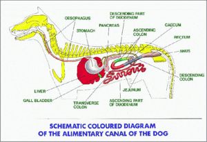
Difference between revisions of "Alimentary System Overview - Anatomy & Physiology"
| Line 60: | Line 60: | ||
==The Avian Digestive Tract== | ==The Avian Digestive Tract== | ||
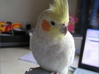
| + | [[Image:Cockatiel.jpg|thumb|right|200px|Cockatiel - Copyright nabrown 2008]] | ||
| + | |||
The [[Avian Digestive Tract - Anatomy & Physiology|avian alimentary system]] differs immensely from the basic mammalian design. Food can move in a retrograde fashion from the [[The Proventriculus - Anatomy & Physiology|proventriculus]] to the [[Crop- Anatomy and Physiology|crop]]. Food can also pass from the [[The Gizzard - Anamtomy & Physiology|gizzard]] back into the [[The Proventriculus - Anatomy & Physiology|proventriculus]] depending on particle size, which is similar to that in mammals. The egestion of bones occurs once the nutritious material has been ingested. During reflux, gastric motility is inhibited and the pellet is expelled through the [[The Avian Oral Cavity - Anatomy & Physiology|oral cavity]] by oesophageal antiperistaltis. This cleans the [[Crop- Anatomy and Physiology|crop]] out and checking the pellet of captive birds should be undertaken daily to assess health. | The [[Avian Digestive Tract - Anatomy & Physiology|avian alimentary system]] differs immensely from the basic mammalian design. Food can move in a retrograde fashion from the [[The Proventriculus - Anatomy & Physiology|proventriculus]] to the [[Crop- Anatomy and Physiology|crop]]. Food can also pass from the [[The Gizzard - Anamtomy & Physiology|gizzard]] back into the [[The Proventriculus - Anatomy & Physiology|proventriculus]] depending on particle size, which is similar to that in mammals. The egestion of bones occurs once the nutritious material has been ingested. During reflux, gastric motility is inhibited and the pellet is expelled through the [[The Avian Oral Cavity - Anatomy & Physiology|oral cavity]] by oesophageal antiperistaltis. This cleans the [[Crop- Anatomy and Physiology|crop]] out and checking the pellet of captive birds should be undertaken daily to assess health. | ||
Revision as of 20:36, 21 June 2010
|
|
Control of Feeding
- Feeding Methods
- Functions of the GIT
- Control of the GIT
- Control of Motility
- Control of GIT Secretions
- Phases of Gastric Secretion
- Neuroendocrine Regulation of Feeding
- The Vomit Reflex
- Mastication
- Deglutition
Oral Cavity
The Oesophagus
Stomach
Small Intestine
Large Intestine
The Liver
The Pancreas
The Peritoneal Cavity
The Avian Digestive Tract
The avian alimentary system differs immensely from the basic mammalian design. Food can move in a retrograde fashion from the proventriculus to the crop. Food can also pass from the gizzard back into the proventriculus depending on particle size, which is similar to that in mammals. The egestion of bones occurs once the nutritious material has been ingested. During reflux, gastric motility is inhibited and the pellet is expelled through the oral cavity by oesophageal antiperistaltis. This cleans the crop out and checking the pellet of captive birds should be undertaken daily to assess health.
The avian intestines shows some species specific anatomical variety, and the hindgut of the avian digestive system differs from mammalian anatomy as it terminates in the cloaca. The external opening through which faecal matter and uric acid is excreted is called the vent. The shape of the vent varies depending on species. Avian species vary in the presence or absence of a gall bladder, and the avian liver differes from mammalian liver being bilobular.
Test yourself with the Alimentary Flashcards
Avian Alimentary Tract Flashcards
References
BOOKS
- Dyce, Sack and Wensing: Textbook of Veterinary Anatomy, 3rd Edition
- Sjaastad, Hove and Sand: Physiology of Domestic Animals, Scandinavian Veterinary Press, Oslo. (735pp)
- Konig and Liebich: Veterinary Anatomy of Domestic Mammals, 3rd Edition
- John E. Harkness and Joseph E. Wagner.: The Biology and Medicine of Rabbits and Rodents, 4th Edition
- Bloom and Fawcett: A Textbook of Histology
- Ian Kay: Introduction to Animal Physiology
- McGeady, Quinn, FitzPatrick, Ryan: Veterinary Embryology, pp. 205-220
- McGavin DM & Zachary, JF: Pathologic Basis of Veterinary Disease, 4th ed, pp. 301-393. Elsevier, St. Louis, Missouri, 2007.
- Reece, WO: Functional Anatomy and Physiology of Domestic Animals, 3rd ed., pp. 312-368. Lippincott Williams & Wilkins, London, England, 2005.
- Young B, Heath, JW: Wheater's Functional Histology: A Text and Colour Atlas, 4th ed, pp. 249-274. Churchill Livingstone, London, England, 2000.
- Andrew A.Mckenzie et al The Capture and Care Manual
- O.Charnock Bradley The Structure of the Fowl, 3rd ed, J.B.Lippincott Company, 1950
IMAGES
- Royal Veterinary College Histology Department
- Dr. Thomas Caceci and Dr. Ihab El-Zhogby Department of Histology, Faculty of Veterinary Medicine, Zagazig University, Egypt
- Nottingham Veterinary School

