Difference between revisions of "Lymphocytes - Introduction"
| (10 intermediate revisions by 5 users not shown) | |||
| Line 1: | Line 1: | ||
| + | {{OpenPagesTop}} | ||
{| align="right" | {| align="right" | ||
|<gallery perrow="1"> | |<gallery perrow="1"> | ||
| − | Image:LH_Lymphocyte_Histology.jpg|<div style="text-align: center;"><p>'''Lymphocyte'''</p><sup> | + | Image:LH_Lymphocyte_Histology.jpg|<div style="text-align: center;"><p>'''Lymphocyte'''</p><sup>Image from [[Blood and Haemopoiesis resource]]</sup></div> |
| − | Image:LH_Lymphocytes_Histology.jpg|<div style="text-align: center;"><p>'''Lymphocytes'''</p><sup> | + | Image:LH_Lymphocytes_Histology.jpg|<div style="text-align: center;"><p>'''Lymphocytes'''</p><sup>Image from [[Blood and Haemopoiesis resource]]</sup></div> |
</gallery> | </gallery> | ||
|} | |} | ||
| Line 10: | Line 11: | ||
==Development== | ==Development== | ||
| − | <p>Both T and B lymphocytes develop from a common stem cell ([[Haematopoiesis - Overview#Colony Forming Units|CFU-L's]]) during [[Leukopoiesis#Lymphopoiesis|lymphopoiesis]].</p> | + | <p>Both T and B lymphocytes develop from a common stem cell ([[Haematopoiesis - Overview#Colony Forming Units|CFU-L's]]) during [[Leukopoiesis#Lymphopoiesis|lymphopoiesis]]. Where they mature accounts for the letter the lymphocytes are given i.e. B cells in the Bone Marrow and T cells in the Thymus. Natural Killer cells develop in the Bone Marrow.</p> |
| − | == | + | ==Lymphocyte Surveillance== |
<p>About two thirds of lymphocytes (all immunocompetent) are circulating in the blood and lymph systems. Most of these are T cells and long lived. In the lymphatic system they survey tissues. The other third do not circulate and are either short lived or immature, or can be specific cells destined for certain tissues i.e. cells that line connective tissue under the epithelium of the respiratory, intestinal and urogential systems.</p> | <p>About two thirds of lymphocytes (all immunocompetent) are circulating in the blood and lymph systems. Most of these are T cells and long lived. In the lymphatic system they survey tissues. The other third do not circulate and are either short lived or immature, or can be specific cells destined for certain tissues i.e. cells that line connective tissue under the epithelium of the respiratory, intestinal and urogential systems.</p> | ||
<p>[[High Endothelial Venules|High endothelial venules (HEV)]] facilitate lymphocyte access to the [[Lymph Nodes - Anatomy & Physiology|lymph nodes]] from the bloodstream. Once inside the lymph node, the naive lymphocytes search for antigens. If there are no antigens present, the naive lymphocytes leave via the efferent lymphatic vessel and return back to the bloodstream. Each lymphocyte can search several [[:Category:Secondary Lymphoid Tissue|secondary lymphoid organs]] each day. This process is called '''surveillance'''.</P> | <p>[[High Endothelial Venules|High endothelial venules (HEV)]] facilitate lymphocyte access to the [[Lymph Nodes - Anatomy & Physiology|lymph nodes]] from the bloodstream. Once inside the lymph node, the naive lymphocytes search for antigens. If there are no antigens present, the naive lymphocytes leave via the efferent lymphatic vessel and return back to the bloodstream. Each lymphocyte can search several [[:Category:Secondary Lymphoid Tissue|secondary lymphoid organs]] each day. This process is called '''surveillance'''.</P> | ||
<p>If a naive lymphocyte recognises an antigen then it differentiates into its adult (mature) form. [[T cell differentiation#Dendritic Cells|Interdigitating dendritic cells]] present antigen to T cells and [[B cell differentiation#Follicular Dendritic Cells|follicular dendritic cells]] present antigen to B cells.</p> | <p>If a naive lymphocyte recognises an antigen then it differentiates into its adult (mature) form. [[T cell differentiation#Dendritic Cells|Interdigitating dendritic cells]] present antigen to T cells and [[B cell differentiation#Follicular Dendritic Cells|follicular dendritic cells]] present antigen to B cells.</p> | ||
<p>B cells proliferate into [[B cell differentiation#Plasma cells|plasma cells]] within sectors of the lymph nodes known as germinal centres, producing antibody.</p> | <p>B cells proliferate into [[B cell differentiation#Plasma cells|plasma cells]] within sectors of the lymph nodes known as germinal centres, producing antibody.</p> | ||
| − | <p>T cells leave the lymph node in '''attack mode''' to locate the infectious organism. The surface molecule L-selectin (which allows the naive lymphocyte to enter the lymph node via an [[Lymph Nodes - Anatomy & Physiology#High endothelial venules|HEV]]) is replaced by the adhesion molecule VLA-4. At the site of inflammation, the VLA-4 receptor recognises VCAM-1 on endothelial cells and the | + | <p>T cells leave the lymph node in '''attack mode''' to locate the infectious organism. The surface molecule L-selectin (which allows the naive lymphocyte to enter the lymph node via an [[Lymph Nodes - Anatomy & Physiology#High endothelial venules|HEV]]) is replaced by the adhesion molecule VLA-4. At the site of inflammation, the VLA-4 receptor recognises VCAM-1 on endothelial cells and the T cell enters the site of disease. [[T cells|CD4<sup>+</sup> T cells]] search for infected macrophages and [[T_cells#Cytotoxic_CD8.2B|CD8<sup>+</sup> T cells]] look for virus infected cells creating an immune response. After the infection has been defeated, memory cells develop which express L-selectin (rather than VLA-4) and continue to search the body in surveillance mode in case the host is re-infected with the disease producing organism. |
| + | {{Template:Learning | ||
| + | |powerpoints = [[Blood and Haemopoiesis resource|Tutorial about histology of blood cells]] | ||
| + | }} | ||
| + | <br><br> | ||
| + | {{Jim Bee 2007}} | ||
| + | {{OpenPages}} | ||
[[Category:Lymphocytes|A]] | [[Category:Lymphocytes|A]] | ||
Latest revision as of 16:34, 2 July 2012
|
Introduction
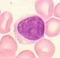
Lymphocytes account for around a third of all circulating leukocytes and are formed in a variety of lymphoid tissues. They are functionally divided into T cells, B cells and Natural Killer (NK) cells. Lymphocytes vary in size (6-30µm) and are classified as small, medium or large. Large cells are either activated lymphocytes or NK cells. The vast majority of circulating lymphocytes are small and of a similar size to erythrocytes. Histologically they are round with a large densely staining nucleus and a thin, often indistinct, rim of cytoplasm. While NK cells can be distinguished by their large granules and kidney shaped nucleus, B and T cells appear the same histologically.
Lymphocytes, along with the associated supporting cells, form the immune system and recognise antigens, produce antibodies and destroy pathogens.
Development
Both T and B lymphocytes develop from a common stem cell (CFU-L's) during lymphopoiesis. Where they mature accounts for the letter the lymphocytes are given i.e. B cells in the Bone Marrow and T cells in the Thymus. Natural Killer cells develop in the Bone Marrow.
Lymphocyte Surveillance
About two thirds of lymphocytes (all immunocompetent) are circulating in the blood and lymph systems. Most of these are T cells and long lived. In the lymphatic system they survey tissues. The other third do not circulate and are either short lived or immature, or can be specific cells destined for certain tissues i.e. cells that line connective tissue under the epithelium of the respiratory, intestinal and urogential systems.
High endothelial venules (HEV) facilitate lymphocyte access to the lymph nodes from the bloodstream. Once inside the lymph node, the naive lymphocytes search for antigens. If there are no antigens present, the naive lymphocytes leave via the efferent lymphatic vessel and return back to the bloodstream. Each lymphocyte can search several secondary lymphoid organs each day. This process is called surveillance.
If a naive lymphocyte recognises an antigen then it differentiates into its adult (mature) form. Interdigitating dendritic cells present antigen to T cells and follicular dendritic cells present antigen to B cells.
B cells proliferate into plasma cells within sectors of the lymph nodes known as germinal centres, producing antibody.
T cells leave the lymph node in attack mode to locate the infectious organism. The surface molecule L-selectin (which allows the naive lymphocyte to enter the lymph node via an HEV) is replaced by the adhesion molecule VLA-4. At the site of inflammation, the VLA-4 receptor recognises VCAM-1 on endothelial cells and the T cell enters the site of disease. CD4+ T cells search for infected macrophages and CD8+ T cells look for virus infected cells creating an immune response. After the infection has been defeated, memory cells develop which express L-selectin (rather than VLA-4) and continue to search the body in surveillance mode in case the host is re-infected with the disease producing organism.
| Lymphocytes - Introduction Learning Resources | |
|---|---|
 Selection of relevant PowerPoint tutorials |
Tutorial about histology of blood cells |
| Originally funded by the RVC Jim Bee Award 2007 |
Error in widget FBRecommend: unable to write file /var/www/wikivet.net/extensions/Widgets/compiled_templates/wrt6633c43ebffb91_66226848 Error in widget google+: unable to write file /var/www/wikivet.net/extensions/Widgets/compiled_templates/wrt6633c43ec55910_19857991 Error in widget TwitterTweet: unable to write file /var/www/wikivet.net/extensions/Widgets/compiled_templates/wrt6633c43ec93fc5_88839448
|
| WikiVet® Introduction - Help WikiVet - Report a Problem |

