Difference between revisions of "Alimentary System - Horse Anatomy"
Ggaitskell (talk | contribs) |
|||
| (2 intermediate revisions by 2 users not shown) | |||
| Line 12: | Line 12: | ||
'''Canines''' | '''Canines''' | ||
| − | :The canines are '''rudimentary''' and are located in the '''diastema'''. The size of the [[ | + | :The canines are '''rudimentary''' and are located in the '''diastema'''. The size of the [[Enamel Organ#Root|root]] is proportionally larger than the [[Enamel Organ#Crown|crown]]. |
'''Molars''' | '''Molars''' | ||
| − | :The molars have enlarged surfaces and higher [[ | + | :The molars have enlarged surfaces and higher [[Enamel Organ#Crown|crowns]]. They have delayed [[Enamel Organ#Root|root]] development and complicated folding of [[Enamel Organ#Enamel|enamel]]. |
'''Incisors''' | '''Incisors''' | ||
| − | :Incisors have high [[ | + | :Incisors have high [[Enamel Organ#Crown|crowns]] and folded [[Enamel Organ#Enamel|enamel]] surfaces. Their [[Enamel Organ#Root|roots]] converge. |
'''Premolars''' | '''Premolars''' | ||
| Line 44: | Line 44: | ||
===Duodenum=== | ===Duodenum=== | ||
[[Image:Section of duodenum from horse.JPG|thumb|right|250px|Section of equine duodenum- © RVC 2008]] | [[Image:Section of duodenum from horse.JPG|thumb|right|250px|Section of equine duodenum- © RVC 2008]] | ||
| − | The descending duodenum is dorsal on the right side of the abdomen, suspended from the dorsal body wall by the mesoduodenum. The mesoduodenum is relatively short, so the duodenum is closely tethered in a constant position. In the right paralumbar fossa region, the descending duodenum turns towards the midline and is attached to the base of the [[Caecum - Anatomy & Physiology|caecum]]. The descending duodenum then runs caudally beneath the [[Liver - Anatomy & Physiology|liver]] to the caudal pole of the right kidney where it has | + | The descending duodenum is dorsal on the right side of the abdomen, suspended from the dorsal body wall by the mesoduodenum. The mesoduodenum is relatively short, so the duodenum is closely tethered in a constant position. In the right paralumbar fossa region, the descending duodenum turns towards the midline and is attached to the base of the [[Caecum - Anatomy & Physiology|caecum]]. The descending duodenum then runs caudally beneath the [[Liver - Anatomy & Physiology|liver]] to the caudal pole of the right kidney where it has it's caudal flexure to become the ascending duodenum. |
===Jejunum=== | ===Jejunum=== | ||
| Line 121: | Line 121: | ||
}} | }} | ||
| − | + | {{review}} | |
| − | + | {{OpenPages}} | |
[[Category:Horse Anatomy]] | [[Category:Horse Anatomy]] | ||
Revision as of 19:05, 19 August 2014
Introduction
The horse is a monogastric hindgut fermenter. The horse evolved for grazing and it does so for up to 17 hours a day. A high proportion of the horse's dietary carbohydrate is in the form of starch. A mature horse eats 2-2.5% of it's body weight in dry matter every day, 1.5-1.75% of this should be fibre (hay/haylage). This is to prevent a rapid drop in pH in the large intestine and also to stimulate peristalsis in the gut and prevent build up of gas.
Oral Cavity
Teeth
Dental Formula
- The formula for deciduous teeth: 2 (I3/3 C0/0 P3/3)
- The formula for permanent teeth: 2 (I3/3 C1/1 P3-4/3 M3/3)
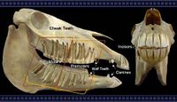
Canines
- The canines are rudimentary and are located in the diastema. The size of the root is proportionally larger than the crown.
Molars
- The molars have enlarged surfaces and higher crowns. They have delayed root development and complicated folding of enamel.
Incisors
Premolars
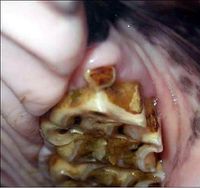
- A horse's Wolf tooth (PM1) is often lacking. Molars and Premolars form a continuous surface. The cheek teeth have a high rate of wear and continually erupt. The upper teeth are wider than the lower. There is no infundibulum in the lower teeth.
Ageing
Horses can be aged by their teeth. At two and a half years of age the first permanent incisor will erupt; at three and a half the second permanent incisor will erupt and at four and a half the third permanent incisor will erupt. Over five years of age the folding of the enamel ring (infundibulum) can indicate age. There is a seven year hook and over 13 years of age a dental star will be present.
The Galvayne's Groove is a brown groove on the upper corner incisor teeth and indicates that the horse is over 10 years old. At 15 the groove will be approximately half way down the tooth; At 20 the groove will run down the whole tooth; Over 20 the groove begins to disappear; At 25 the groove will only be visible on the bottom half of the tooth. At 30 the groove will usually be gone.
Palate
Horses have a tight laryngeal cuff around the laryngeal entrance, therefore the soft palate cannot be raised for long periods of time. This causes them to be obligate nasal breathers. Laryngeal cuffing also prevents vomiting. There are no specific species diferences in the hard palate.
Oesophagus
In the horse, the oesophageal lumen narrows at the thoracic inlet and oesophageal hiatus of the diaphragm; this predisposes them to impaction (choke). Another factor specific to horses is that striated muscle exists only in the rostral 65% of the oesophagus.
Stomach
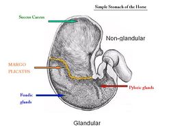
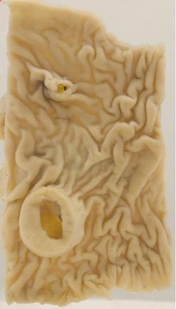
The horse has a monogastric stomach located on the left side of the abdomen. A region called the margo plicatus is present which separates the glandular and non-glandular parts of the equine stomach. The non-glandular area is lined with squamous epithelium (not columnar).
The stomach is relatively small (10% GIT) and its capacity is 8-16 litres. The equine stomach is rarely empty, retention time is short and expulsion into the duodenum stops when feeding stops. Although fluid exits quickly, feed particles can be retained for more than 48 hours as digestion is initiated in the stomach. A 500kg horse can produce 30 litres of gastric juice in 24 hours. The strong cardiac sphincter allows movement of gas and fluid into the stomach, but not out of it. This prevents the animal from vomiting. Therefore, any disorder that results in aboral fluid movement from the small intestine results in fluid accumulation in the stomach (gastric reflux), dilation and eventually gastric rupture if left untreated.
Small Intestine
Duodenum
The descending duodenum is dorsal on the right side of the abdomen, suspended from the dorsal body wall by the mesoduodenum. The mesoduodenum is relatively short, so the duodenum is closely tethered in a constant position. In the right paralumbar fossa region, the descending duodenum turns towards the midline and is attached to the base of the caecum. The descending duodenum then runs caudally beneath the liver to the caudal pole of the right kidney where it has it's caudal flexure to become the ascending duodenum.
Jejunum
The jejunum is confined to the left dorsal part of the abdomen associated with a long mesentery. It is restricted to this position by the large caecum on the right, and ascending colon ventrally on both sides.
Ileum
The terminal portion of the small intestine is the ileum, which joins the caecum at its dorso-medial aspect. The ileal mesentery attaches to the caecum at the dorsal caecal band.
Large Intestine
Undigested material spends a long time in the caecum and large intestine undergoing microbial fermentation, mainly of cellulose (95% after 65 hours).
In the hindgut of the horse; 75-85% of insoluble carbohydrates is digested, 15-30% of soluble carbohydrates and 30% of protein is digested. A lot of absorption of volatile fatty acids (VFAs) and water occurs in the large intestine which pass readily into the blood. Electrolytes are also absorbed in the large intestine; 95% of sodium and chloride and 75% of potassium and phosphate. To mix the contents of the large intestines, the taenia and circular muscle of the tunica muscularis contract. This also transports the ingesta through the large intestine and brings the products of fermentation in contact with the epithelium.
Caecum
The caecum is the main site of microbial fermentation, followed by the ascending then descending colons. It is located on the right side of the abdomen. It is very large, roughly 1m in length with a 30L capacity. It consists of a base, body and apex (blind ending). The base lies in the right dorsal part of the abdomen, in contact with the abdominal roof. The apex lies on the ventral abdominal wall, and terminates at the level of the xiphoid cartilage. It exists at the junction with the ileum and colon.
The caecocolic orifice is where the caecum opens into the ascending colon. This exists as a transverse slit formed by a constriction of the ascending colon. There is a sphincter at this point which prevents backward flow of ingesta when the colon contracts.
The ileum opens into the caecum at the ileal papilla. This is a small projection into the caecum housing the ileal sphincter and venous plexus that, together, control the ileal orifice.
Taeniae are present. Taeniae are formed by concentration of the longitudinal muscle layer. Between the taeniae are sacculations, or haustra. Haustra appear as folds on the interior surface. There are four taeniae over the caecum; dorsal, ventral, lateral and medial. The dorsal taenia provides the attachment site for the ileocaecal fold, which joins the caecum to the ileum.
The lateral taenia provides the attachment site for the caecocolic fold, which joins the caecum to the ascending colon. The ventral taenia is free.
The medial and lateral taeniae are where the caecal vessels and lymph nodes are located. Ingesta is regularly transported from the ileum to the caecum, this movement can be heard upon auscultation of the right dorsal quadrant of the caudal abdomen. Ausculatation of this area is carried out in the assessment of colic. In the horse, the caecum is responsible for the digestion of complex carbohydrates such as cellulose.
Colon
Ascending Colon
The ascending colon is very large and takes up most of the ventral abdomen. It is the shape of a double "U", where one "U" is on top of the other. There are four limbs that lie parallel to each other, and three flexures that change these direction of the limbs.
The sequence of the limbs and flexures of the ascending colon is as follows; Right Ventral Colon (for those with an RVC bias remember, "the RVC comes first!"), passes out of the caecocolic orifice on the right side of the abdomen and continues cranially to the xiphoid region; Sternal Flexure, passes across the midline from right to left, Left Ventral Colon, runs caudally on the left ventral abdominal floor; Pelvic Flexure, turns dorsally just cranial to the pelvic inlet and then runs cranially to the diaphragm, Left Dorsal Colon, runs cranially, parallel and dorsal to the left ventral colon; Diaphragmatic Flexure, turns caudally at the diaphragm; Right Dorsal Colon, continues caudally on the right. It is the shortest limb of the ascending colon.
The transverse colon continues on from the right dorsal colon as the right dorsal colon turns medially. The right dorsal colon is attached by a mesentery to the dorsal abdominal wall, the base of the caecum, the root of the mesentery and the pancreas. This anatomical arrangement of mesentery allows the left ascending colon to twist and is a common cause of colic (colonic torsion).
The ventral parts of the ascending colon are attached to the dorsal parts by a short mesocolon. The mesocolon houses the blood vessels, nerves and lymphatics. In the ventral colon many important digestive and absorptive functions take place, whilst the dorsal colon is mainly responsible for transportation of ingesta. Taeniae are present. Different parts of the colon can be distinguished by the number of taeniae present:
The right and left ventral colon and the sternal flexure have four taeniae. The left dorsal colon and pelvic flexure have one taenia and the right dorsal colon and diaphragmatic flexure have three taeniae.
Transverse Colon
The transverse colon is short. It passes from across the midline from right to left. It passes cranial to the root of the mesentery. It has two taeniae. It turns caudally to become the descending colon at the level of the left kidney.
Descending Colon
The descending colon is between 2-4 meters long. It is suspended by a long mesentery; mesocolon descendens. The descending colon has two taeniae. Between the two taeniae are distinct sacculations that house the faecal balls.
Rectal Palpation
Rectal palpation is a useful technique and is often used to assess colic. Structures that can be palpated per rectum include; faecal balls in the descending colon, the bladder, the reproductive organs in the mare, the base of the caecum, the root of the mesentery, the left kidney, +/- the nephrosplenic ligament, the left dorsal colon and the pelvic flexure of the ascending colon. NB: This is a common site of impaction.
Microbial Environment
Microbes convert carbohydrates to volatile fatty acids (VFAs). The horse receives 75% of its energy requirements from VFAs. The large intestine is buffered by the secretion of large amounts of bicarbonate from the pancreas and the ileum. Glands in the wall of the large intestine may also produce bicarbonate. The microbial population exists in the caecum and ventral colon.
The environment is mixed; there are both bacteria and protozoa. Microbes are anaerobic. The microbial population is dependent on diet and frequency of feeding, as different microbes are suited to digesting different particles. The number of microbes can change 100 fold in a 24 hour period. VFAs produced are absorbed across the intestinal wall. Urea from the blood is transported to the intestinal lumen to be used by microbes, which also use nitrogen from the diet. Environmental factors of the caecum and ventral colon can influence fermentation of microbial population.
Environmental factors include: frequent intake of food, constant temperature, constant mixing, removal of the products of fermentation by absorption and peristalsis and the stable osmotic environment i.e. normal intake of water.
VFA's produced include Acetate, Propionate and Butyrate. Factors that promote VFA production include an optimum pH of 6.5, an anaerobic environment and gut motility.
Liver
The liver is contained entirely within the rib cage, to the right of the midline. It is less lobated than in other species. The larger right lobe is undivided, and the left lobe subdivided. The caudate lobe is notched at the ventral free border. There is no papillary process. Horses have no gall bladder, and the hepatic ducts are relatively wide as a result. In the foal, the liver is larger and more symmetrical. The bile duct opens into the duodenum at the same papillae as the major pancreatic duct. Bile is constantly secreted.
Pancreas
The pancreas lies mainly on the right, in the very dorsal part of the abdomen. It is triangular in shape and lies within the sigmoid flexure of the duodenum. The lobes are less distinguishable compared to the dog. The ventral surface is directly attached to the right dorsal colon and base of the caecum. The dorsal surface is directly attached to the right kidney and liver. The portal vein perforates the pancreas at the pancreatic ring. Both the pancreatic and accessory ducts persist throughout development. There is a constant secretion of pancreatic juice, which increases after feeding. This provides the caecum and colon with a constant supply of buffered solution, which maintains a stable environment important for microbe survival.
| Alimentary System - Horse Anatomy Learning Resources | |
|---|---|
 Test your knowledge using flashcard type questions |
Horse digestive system |
 Selection of relevant videos |
Foal gastrointestinal tract potcast Abdominal viscera of the horse dissection Equine left-sided abdominal and thoracic topography dissection Equine left-sided abdominal and thoracic topography dissection 2 Equine stomach potcast |
Anatomy Museum Resources |
PowerPoint covering the anatomy and physiology of the equine head and dentition, including the skeletal aspects, the physiology of mastication and it’s associated anatomy as well as common dental abnormalities. Equine Caecum and Colon - Left View Equine Caecum and Colon - Right View Equine Oesophagus Histology Equine Duodenum Histology 1 Equine Duodenum Histology 2 Smooth Muscle Histology of Equine Duodenum |
| This article has been peer reviewed but is awaiting expert review. If you would like to help with this, please see more information about expert reviewing. |
Error in widget FBRecommend: unable to write file /var/www/wikivet.net/extensions/Widgets/compiled_templates/wrt69a2002491e3c6_08758118 Error in widget google+: unable to write file /var/www/wikivet.net/extensions/Widgets/compiled_templates/wrt69a200249ff609_02087197 Error in widget TwitterTweet: unable to write file /var/www/wikivet.net/extensions/Widgets/compiled_templates/wrt69a20024aa19e1_94052918
|
| WikiVet® Introduction - Help WikiVet - Report a Problem |


