Difference between revisions of "Alimentary System Overview - Anatomy & Physiology"
Fiorecastro (talk | contribs) |
|||
| (167 intermediate revisions by 9 users not shown) | |||
| Line 1: | Line 1: | ||
| − | |||
| − | |||
| − | |||
| − | |||
| − | |||
| − | |||
| − | |||
| − | |||
| − | |||
| − | |||
| − | |||
| − | |||
| − | |||
| − | |||
| − | |||
| − | |||
| − | |||
| − | |||
| − | |||
| − | |||
| − | |||
| − | |||
| − | |||
| − | |||
| − | |||
| − | |||
| − | |||
| − | |||
| − | |||
| − | |||
| − | |||
| − | |||
| − | |||
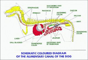
| + | [[Image:Alimentary Canine.jpg|thumb|right|300px|The Alimentary Tract (Canine) - Copyright Prof. Pat Mccarthy]] | ||
==Introduction== | ==Introduction== | ||
| + | The anatomy of the alimentary system begins rostrally with the [[Oral Cavity Overview - Anatomy & Physiology|oral cavity]], which is the first section of the alimentary tract that receives food. It provides the digestive functions of prehension, [[Mastication|mastication]] and insalivation and also plays a role in the respiratory system through oral breathing when the nasopharynx is impaired. The oral cavity or mouth, includes accessory structures - [[Salivary Glands - Anatomy & Physiology|the salivary glands]], projecting structures - [[:Category:Teeth - Anatomy & Physiology|the teeth]] and [[Tongue - Anatomy & Physiology|tongue]], and the boundaries enclosing the oral cavity; the [[Lips|lips]], [[Cheeks|cheeks]], [[Soft Palate|soft]] and [[Hard Palate|hard palates]], and the [[Oropharynx - Anatomy & Physiology|oropharynx]]. In anatomical terms, the oropharynx is common to both the alimentary and the respiratory system, and the hard and soft palate forms the boundary between the oral and nasal cavities in many species. | ||
| − | + | Food passes from the oral cavity into the [[oesophagus - Anatomy & Physiology|oesophagus]] and from here to the stomach. In evolutionary terms, various adaptations to the anatomy of the stomach reflect the digestive needs of the species based on their natural diet. The [[Ruminant Stomach - Anatomy & Physiology|ruminant stomach]] for example, is composed of 4 separate compartments; the rumen, the reticulum, the omasum and the abomasum. The first three compartments are adapted to digest complex carbohydrates with the aid of microorganisms which produce [[Volatile Fatty Acids|volatile fatty acids]] - the major energy source of ruminants. The last compartment, the abomasum resembles the simple [[Monogastric Stomach - Anatomy & Physiology|monogastric stomach]] of a carnivore in structure and function. As a further adaptation, the [[Oesophageal Groove|oesophageal groove]] is present in newborn ruminants; it is a channel which directs milk from the oesophagus into the rumen, omasum and then abomasum, bypassing the reticulum. | |
| − | |||
| − | |||
| − | |||
| − | |||
| − | |||
| − | |||
| − | + | The stomach passes into the [[Small Intestine Overview - Anatomy & Physiology|small intestine]], which is subdivided into three sections; the [[Duodenum - Anatomy & Physiology|duodenum]], the [[Jejunum - Anatomy & Physiology|jejunum]] and the [[Ileum - Anatomy & Physiology|ileum]]. The small intestine receives the ingested food from the stomach and is the main site of the chemical degradation and absorption of ingesta. Fats are exclusively broken down in this part of the alimentary tract. Carbohydrates and proteins that are not degraded in the small intestine are available for microbial fermentation in the large intestine. The wall of the small intestine produces enzymes for the digestion of protein, carbohydrate and fat. The [[Pancreas - Anatomy & Physiology|pancreas]] also produces digestive enzymes to aid this process. The [[Gall Bladder - Anatomy & Physiology|gall bladder]] stores bile which is produced in the [[Liver - Anatomy & Physiology|liver]] and emulsifies fats for digestion. Absorption in the small intestine is facilitated by ridges in the small intestine and by the presence of villi and microvilli. | |
| − | + | The [[Large Intestine - Anatomy & Physiology|large intestine]] begins at the [[Caecum - Anatomy & Physiology|caecum]], and includes the [[Colon - Anatomy & Physiology|colon]], the [[Rectum - Anatomy & Physiology|rectum]] and the [[Anus - Anatomy & Physiology|anus]]. Water, electrolytes and nutrients are absorbed which concentrates the ingesta into faeces. There is no secretion of enzymes and any digestion that takes place is carried out by microbes. All species have a large microbial population living in the large intestine, which is of particular importance to the [[Hindgut Fermenters - Anatomy & Physiology|hindgut fermenters]] such as the horse. For this reason, hindgut fermenters have a more complex large intestine with highly specialised regions for fermentation. The volatile fatty acid products of microbial fermentation are absorbed in the colon. | |
| − | |||
| − | |||
| − | |||
| − | |||
| − | |||
| − | |||
| − | + | The stomach, small intestines and large intestines are situatied within the abdominal or [[Peritoneal Cavity - Anatomy & Physiology|peritoneal cavity]]. The peritoneum is the serous membrane that lines the abdominal cavity; it produces fluid to lubricate abdominal viscera and enhances the immune response and walls off infection in the abdomen to prevent peritonitis. | |
| − | + | ||
| − | + | *[[Camelid Stomach - Anatomy & Physiology|The Camelid Stomach]] | |
| − | |||
| − | |||
| − | |||
| − | [[ | ||
| − | == | + | ==The Physiology of feeding== |
| + | Different hormones, neurotransmitters and reflexes are involved in the complicated [[Control of Feeding - Anatomy & Physiology|process of feeding]] in animals. [[Control of Feeding - Anatomy & Physiology#Control of GIT Secretions|Secretions]] and [[Control of Feeding - Anatomy & Physiology#Control of Motility|motility]] of the gastrointestinal tract are stimulated and carefully regulated by numerous factors, including environmental stimuli and the presence of food in different parts of the gastrointestinal tract, which is detected by chemoreceptors and mechanical receptors. Motility is modified by both intrinsic and extrinsic nervous sytems, and neurological reflex mechanisms prevent food from accidentally passing into the trachea during [[Deglutition|deglutition]], or swallowing. | ||
| − | + | When a harmful substance is ingested the body acts to eliminate it in different ways to prevent the animal becoming ill, for example, through [[Control of Feeding - Anatomy & Physiology#The Vomit Reflex|vomiting]] or diarrhoea. If one or more of the [[Control of Feeding - Anatomy & Physiology#Neuroendocrine Regulation of Feeding|neuroendocrine pathways]] involved with the control of feeding is damaged or inhibited, then problems such as obesity can occur. | |
| − | |||
| − | |||
| − | |||
| − | + | There are many species differences in the phsiology of feeding, from different [[Control of Feeding - Anatomy & Physiology#Feeding Methods|feeding methods]] to adaptations during the digestive process such as additional cycles of [[Mastication|mastication]] which is seen during [[Rumination|rumination]] and [[Eructation|eructation]]. | |
| − | The | + | ==The Avian Digestive Tract== |
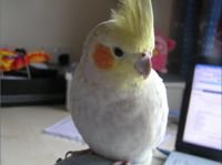
| + | [[Image:Cockatiel.jpg|thumb|right|200px|Cockatiel - Copyright nabrown 2008]] | ||
| − | + | The [[Avian Digestive Tract - Anatomy & Physiology|avian alimentary system]] differs immensely from the basic mammalian design. Food can move in a retrograde fashion from the [[Proventriculus - Anatomy & Physiology|proventriculus]] to the [[Crop - Anatomy and Physiology|crop]]. Food can also pass from the [[Gizzard - Anatomy & Physiology|gizzard]], which is the equivalent of a muscular stomach back into the [[Proventriculus - Anatomy & Physiology|proventriculus]], or glandular stomach depending on particle size. The egestion of bones occurs once the nutritious material has been ingested. During reflux, gastric motility is inhibited and the pellet is expelled through the [[Avian Oral Cavity - Anatomy & Physiology|oral cavity]] by oesophageal antiperistaltis. This cleans the [[Crop - Anatomy and Physiology|crop]] out and checking the pellet of captive birds should be undertaken daily to assess health. | |
| − | |||
| − | |||
| − | |||
| − | |||
| − | |||
| − | |||
| − | |||
| − | |||
| − | + | The [[Avian Intestines - Anatomy & Physiology|avian intestines]] shows some species specific anatomical variety, and the hindgut of the avian digestive system differs from mammalian anatomy as it terminates in the [[Avian Vent and Cloaca - Anatomy & Physiology|cloaca]]. The external opening through which faecal matter and uric acid is excreted is called the [[Avian Vent and Cloaca - Anatomy & Physiology|vent]]. The shape of the vent varies depending on species. Avian species vary in the presence or absence of a [[Avian_Liver_- Anatomy & Physiology#Gallbladder:_Species_Differences|gall bladder]], and the avian [[Avian Liver - Anatomy & Physiology|liver]] differs from the mammalian liver, being bilobular. | |
| − | + | ==Test yourself with the Alimentary Flashcards== | |
| − | + | [[:Category:Alimentary System Anatomy & Physiology Flashcards|Alimentary Flashcards]] | |
| − | |||
| − | |||
| − | + | [[The Avian Alimentary Tract - Anatomy & Physiology - Flashcards|Avian Alimentary Tract Flashcards]] | |
| + | ==References== | ||
| + | BOOKS | ||
| + | *Dyce, Sack and Wensing: Textbook of Veterinary Anatomy, 3rd Edition | ||
| + | *Sjaastad, Hove and Sand: Physiology of Domestic Animals, Scandinavian Veterinary Press, Oslo. (735pp) | ||
| + | *Konig and Liebich: Veterinary Anatomy of Domestic Mammals, 3rd Edition | ||
| + | *John E. Harkness and Joseph E. Wagner.: The Biology and Medicine of Rabbits and Rodents, 4th Edition | ||
| + | *Bloom and Fawcett: A Textbook of Histology | ||
| + | *Ian Kay: Introduction to Animal Physiology | ||
| + | *McGeady, Quinn, FitzPatrick, Ryan: Veterinary Embryology, pp. 205-220 | ||
| + | *McGavin DM & Zachary, JF: Pathologic Basis of Veterinary Disease, 4th ed, pp. 301-393. Elsevier, St. Louis, Missouri, 2007. | ||
| + | *Reece, WO: Functional Anatomy and Physiology of Domestic Animals, 3rd ed., pp. 312-368. Lippincott Williams & Wilkins, London, England, 2005. | ||
| + | *Young B, Heath, JW: Wheater's Functional Histology: A Text and Colour Atlas, 4th ed, pp. 249-274. Churchill Livingstone, London, England, 2000. | ||
| + | *Andrew A.Mckenzie et al The Capture and Care Manual | ||
| + | *O.Charnock Bradley The Structure of the Fowl, 3rd ed, J.B.Lippincott Company, 1950 | ||
| − | + | IMAGES | |
| + | *Royal Veterinary College Histology Department | ||
| + | *Dr. Thomas Caceci and Dr. Ihab El-Zhogby Department of Histology, Faculty of Veterinary Medicine, Zagazig University, Egypt | ||
| + | *Nottingham Veterinary School | ||
| − | |||
| − | + | [[Category:Alimentary System - Anatomy & Physiology]] | |
| − | |||
Latest revision as of 15:19, 4 January 2023
Introduction
The anatomy of the alimentary system begins rostrally with the oral cavity, which is the first section of the alimentary tract that receives food. It provides the digestive functions of prehension, mastication and insalivation and also plays a role in the respiratory system through oral breathing when the nasopharynx is impaired. The oral cavity or mouth, includes accessory structures - the salivary glands, projecting structures - the teeth and tongue, and the boundaries enclosing the oral cavity; the lips, cheeks, soft and hard palates, and the oropharynx. In anatomical terms, the oropharynx is common to both the alimentary and the respiratory system, and the hard and soft palate forms the boundary between the oral and nasal cavities in many species.
Food passes from the oral cavity into the oesophagus and from here to the stomach. In evolutionary terms, various adaptations to the anatomy of the stomach reflect the digestive needs of the species based on their natural diet. The ruminant stomach for example, is composed of 4 separate compartments; the rumen, the reticulum, the omasum and the abomasum. The first three compartments are adapted to digest complex carbohydrates with the aid of microorganisms which produce volatile fatty acids - the major energy source of ruminants. The last compartment, the abomasum resembles the simple monogastric stomach of a carnivore in structure and function. As a further adaptation, the oesophageal groove is present in newborn ruminants; it is a channel which directs milk from the oesophagus into the rumen, omasum and then abomasum, bypassing the reticulum.
The stomach passes into the small intestine, which is subdivided into three sections; the duodenum, the jejunum and the ileum. The small intestine receives the ingested food from the stomach and is the main site of the chemical degradation and absorption of ingesta. Fats are exclusively broken down in this part of the alimentary tract. Carbohydrates and proteins that are not degraded in the small intestine are available for microbial fermentation in the large intestine. The wall of the small intestine produces enzymes for the digestion of protein, carbohydrate and fat. The pancreas also produces digestive enzymes to aid this process. The gall bladder stores bile which is produced in the liver and emulsifies fats for digestion. Absorption in the small intestine is facilitated by ridges in the small intestine and by the presence of villi and microvilli.
The large intestine begins at the caecum, and includes the colon, the rectum and the anus. Water, electrolytes and nutrients are absorbed which concentrates the ingesta into faeces. There is no secretion of enzymes and any digestion that takes place is carried out by microbes. All species have a large microbial population living in the large intestine, which is of particular importance to the hindgut fermenters such as the horse. For this reason, hindgut fermenters have a more complex large intestine with highly specialised regions for fermentation. The volatile fatty acid products of microbial fermentation are absorbed in the colon.
The stomach, small intestines and large intestines are situatied within the abdominal or peritoneal cavity. The peritoneum is the serous membrane that lines the abdominal cavity; it produces fluid to lubricate abdominal viscera and enhances the immune response and walls off infection in the abdomen to prevent peritonitis.
The Physiology of feeding
Different hormones, neurotransmitters and reflexes are involved in the complicated process of feeding in animals. Secretions and motility of the gastrointestinal tract are stimulated and carefully regulated by numerous factors, including environmental stimuli and the presence of food in different parts of the gastrointestinal tract, which is detected by chemoreceptors and mechanical receptors. Motility is modified by both intrinsic and extrinsic nervous sytems, and neurological reflex mechanisms prevent food from accidentally passing into the trachea during deglutition, or swallowing.
When a harmful substance is ingested the body acts to eliminate it in different ways to prevent the animal becoming ill, for example, through vomiting or diarrhoea. If one or more of the neuroendocrine pathways involved with the control of feeding is damaged or inhibited, then problems such as obesity can occur.
There are many species differences in the phsiology of feeding, from different feeding methods to adaptations during the digestive process such as additional cycles of mastication which is seen during rumination and eructation.
The Avian Digestive Tract
The avian alimentary system differs immensely from the basic mammalian design. Food can move in a retrograde fashion from the proventriculus to the crop. Food can also pass from the gizzard, which is the equivalent of a muscular stomach back into the proventriculus, or glandular stomach depending on particle size. The egestion of bones occurs once the nutritious material has been ingested. During reflux, gastric motility is inhibited and the pellet is expelled through the oral cavity by oesophageal antiperistaltis. This cleans the crop out and checking the pellet of captive birds should be undertaken daily to assess health.
The avian intestines shows some species specific anatomical variety, and the hindgut of the avian digestive system differs from mammalian anatomy as it terminates in the cloaca. The external opening through which faecal matter and uric acid is excreted is called the vent. The shape of the vent varies depending on species. Avian species vary in the presence or absence of a gall bladder, and the avian liver differs from the mammalian liver, being bilobular.
Test yourself with the Alimentary Flashcards
Avian Alimentary Tract Flashcards
References
BOOKS
- Dyce, Sack and Wensing: Textbook of Veterinary Anatomy, 3rd Edition
- Sjaastad, Hove and Sand: Physiology of Domestic Animals, Scandinavian Veterinary Press, Oslo. (735pp)
- Konig and Liebich: Veterinary Anatomy of Domestic Mammals, 3rd Edition
- John E. Harkness and Joseph E. Wagner.: The Biology and Medicine of Rabbits and Rodents, 4th Edition
- Bloom and Fawcett: A Textbook of Histology
- Ian Kay: Introduction to Animal Physiology
- McGeady, Quinn, FitzPatrick, Ryan: Veterinary Embryology, pp. 205-220
- McGavin DM & Zachary, JF: Pathologic Basis of Veterinary Disease, 4th ed, pp. 301-393. Elsevier, St. Louis, Missouri, 2007.
- Reece, WO: Functional Anatomy and Physiology of Domestic Animals, 3rd ed., pp. 312-368. Lippincott Williams & Wilkins, London, England, 2005.
- Young B, Heath, JW: Wheater's Functional Histology: A Text and Colour Atlas, 4th ed, pp. 249-274. Churchill Livingstone, London, England, 2000.
- Andrew A.Mckenzie et al The Capture and Care Manual
- O.Charnock Bradley The Structure of the Fowl, 3rd ed, J.B.Lippincott Company, 1950
IMAGES
- Royal Veterinary College Histology Department
- Dr. Thomas Caceci and Dr. Ihab El-Zhogby Department of Histology, Faculty of Veterinary Medicine, Zagazig University, Egypt
- Nottingham Veterinary School

